Shows
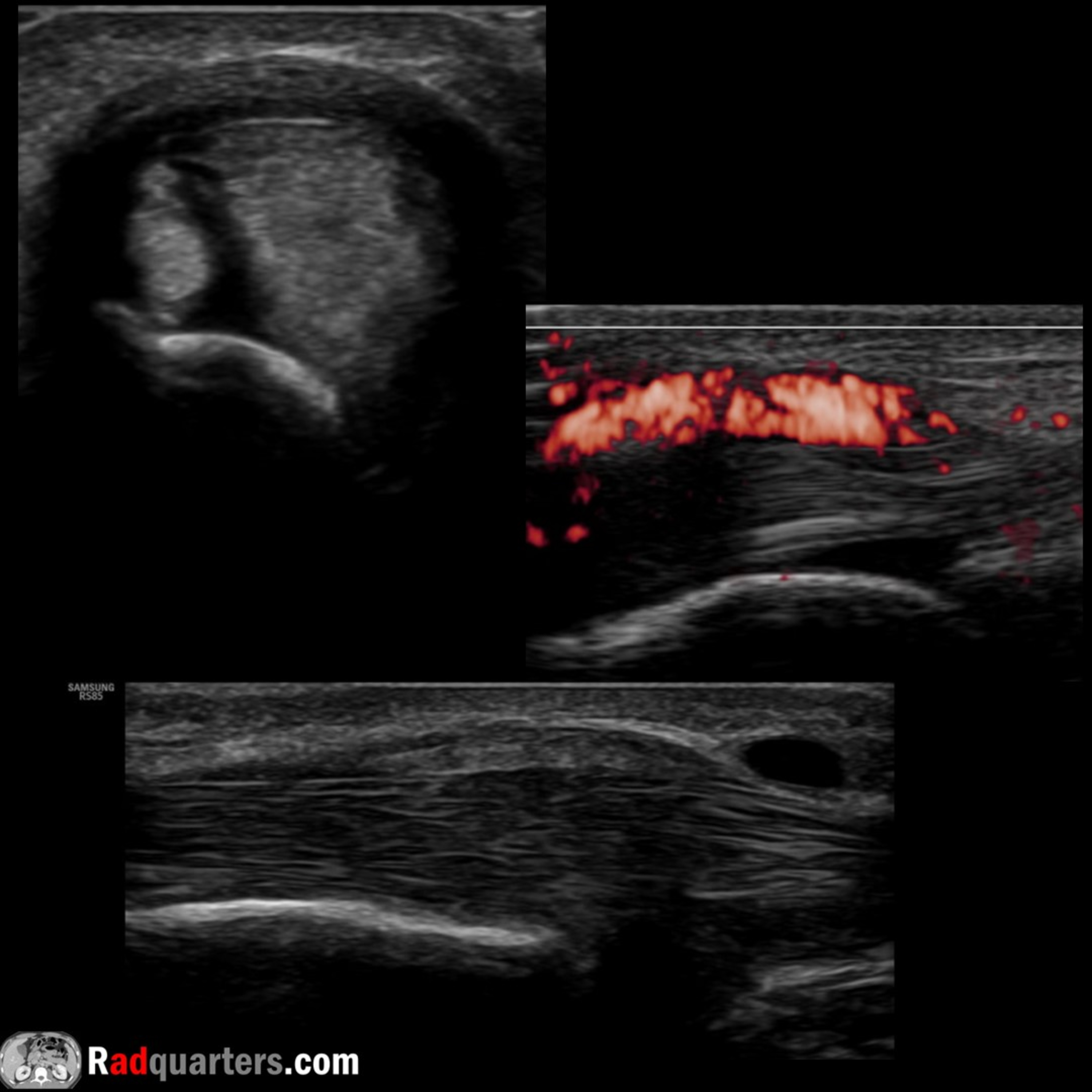
 Radiology Lectures | RadquartersUltrasound of de Quervain’s TenosynovitisIn this radiology lecture, we review the ultrasound appearance of de Quervain’s Tenosynovitis!
Key teaching points include:
Stenosing tenosynovitis of first extensor compartment tendons = Extensor pollicis brevis (EPB) and abductor pollicis longus (APL)
Second most common hand entrapment tendinopathy after trigger finger
Most common in middle-aged females
Associations include repetitive hand motions, pregnancy, arthritis, and trauma
Clinical presentation: Pain with thumb and wrist movement, tenderness and swelling at radial styloid
Positive Finkelstein maneuver may be present: Grasp thumb, ulnar deviate hand = Pain over distal radius
Ultrasound findings: Increased fluid in EPB/APL tendon sheath (tenosynovitis), hy...2024-09-1208 min
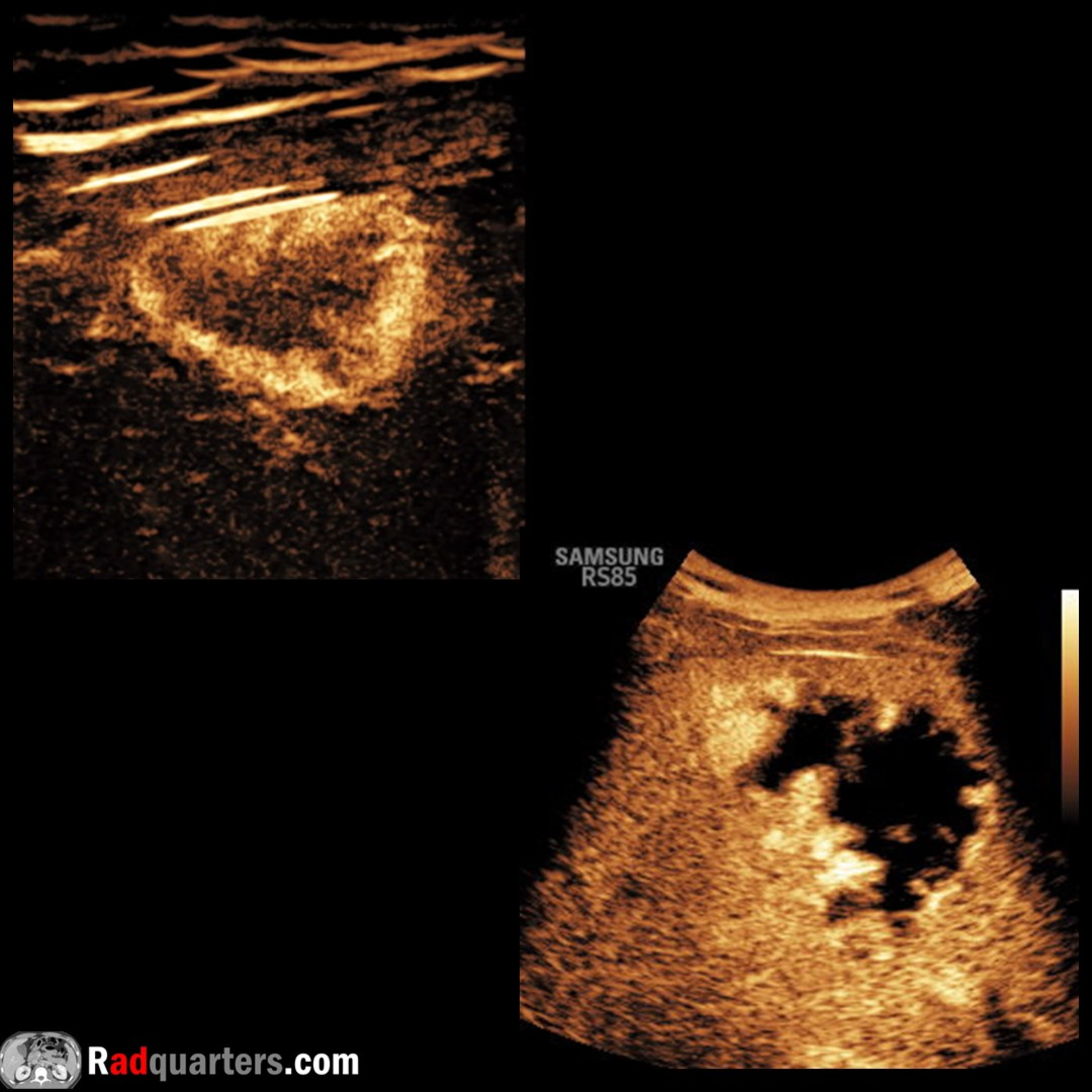
 Radiology Lectures | RadquartersContrast-Enhanced Ultrasound of HemangiomaIn this radiology lecture, we review the contrast-enhanced ultrasound appearance of hepatic hemangioma!
Key teaching points include:
Microbubble contrast agents are gas-filled microspheres with a lipid or protein shell
Sulfur hexafluoride lipid-type A microspheres: Inert gas of six fluoride atoms bound to one sulfur atom, surrounded by a phospholipid shell
Similar in size to red blood cells, unique when compared to the molecular sizes of CT and MR imaging contrast agents. Small enough to cross capillary beds, too large to enter interstitial space = Pure intravascular agents, ideal for assessing vascularity and perfusion
After IV injection, US...2024-08-1510 min
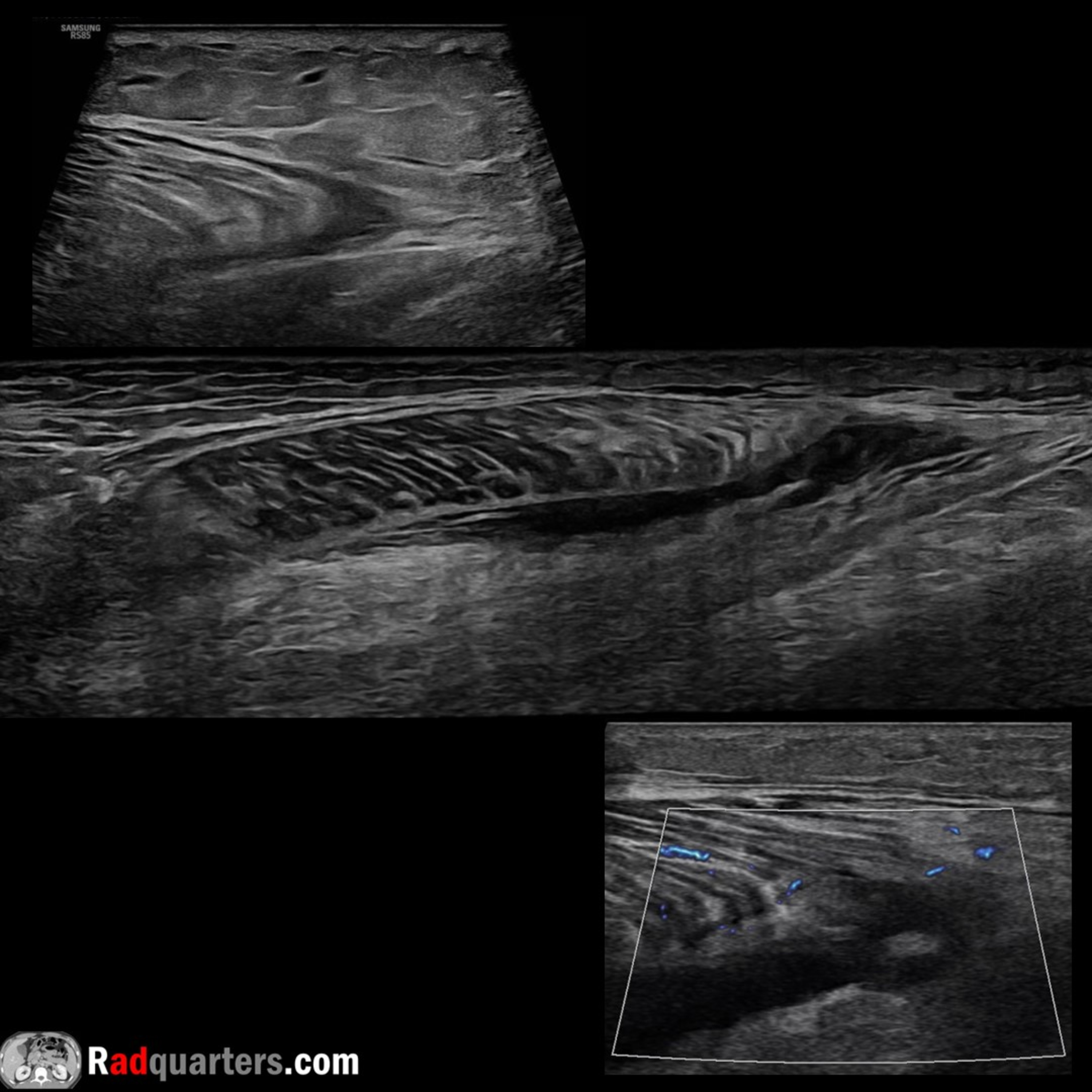
 Radiology Lectures | RadquartersUltrasound of Tennis LegIn this radiology lecture, we review the ultrasound appearance of tennis leg, including medial gastrocnemius and plantaris injury!
Key teaching points include:
Tennis leg = Injury to muscles of the calf. Tear of myotendinous junction of medial head of gastrocnemius, rupture of plantaris tendon (less common), in isolation or together
Classically described in tennis players, but can occur in various athletic activities (running, skiing) with extension of knee and forced dorsiflexion of ankle. Typically seen in middle-aged, active individuals
Clinical: Sudden sharp calf pain with associated popping/snapping sensation followed by tenderness and swelling
Gastrocnemius & soleus are...2024-07-1409 min
 Radiology Lectures | RadquartersUltrasound of Interstitial Ectopic PregnancyIn this radiology lecture, we review the ultrasound appearance of interstitial ectopic pregnancy!
Key teaching points include:
Interstitial ectopic pregnancies are rare, occurring in proximal (interstitial) portion of fallopian tube within muscle wall of uterus
Much less common than tubal ectopic pregnancy occurring in the more distal ampullary and isthmic portions of fallopian tube
Interstitial ectopic pregnancies are important because higher morbidity and mortality due to later presentation and risk of life-threatening hemorrhage
Abnormally eccentric gestational sac with thin surrounding myometrium: less than 5 mm myometrial thickness highly suspicious
“Interstitial line” sign: Thin echogenic line extending from endo...2024-06-0607 min
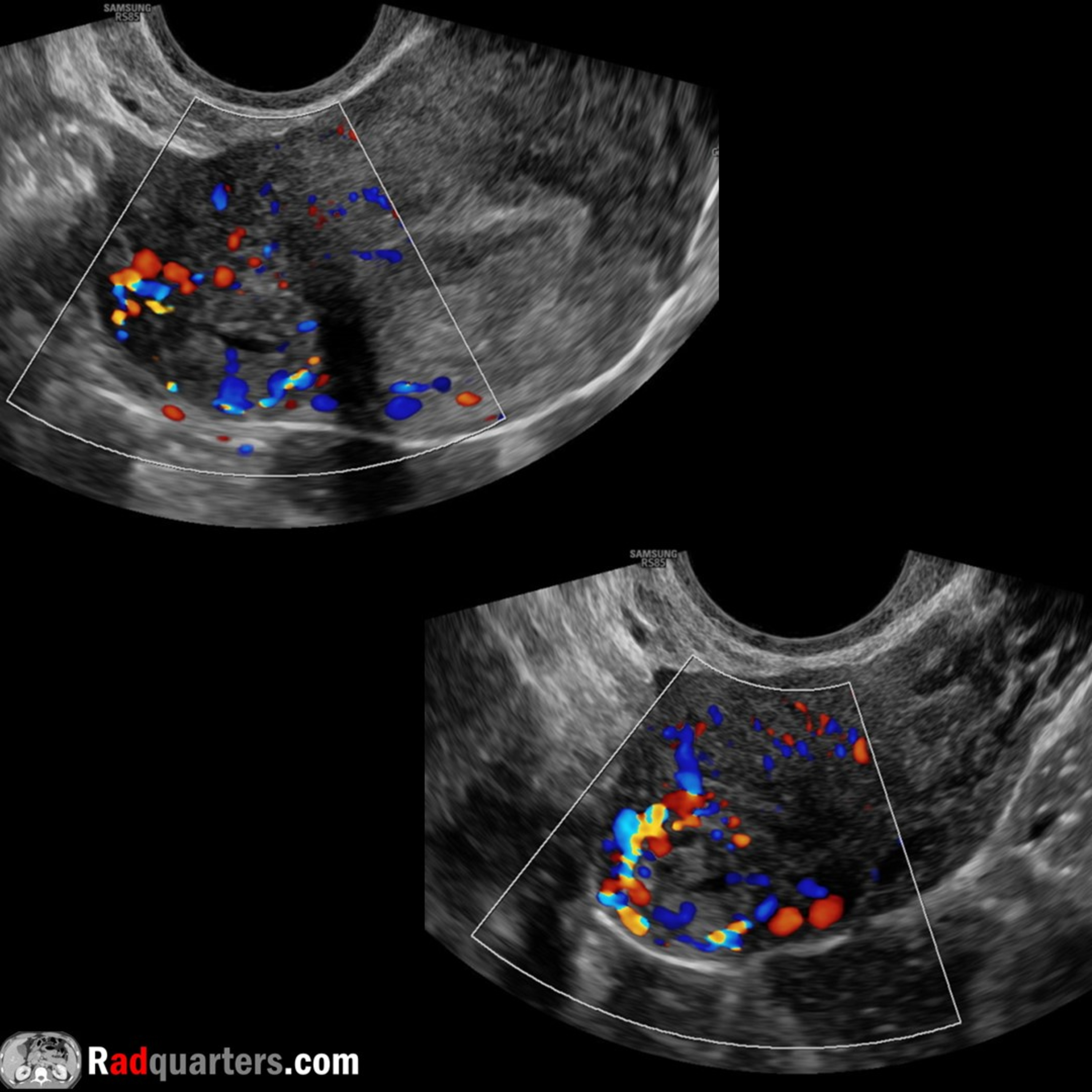
 Radiology Lectures | RadquartersUltrasound of Ovarian Serous CystadenocarcinomaIn this radiology lecture, we review the ultrasound appearance of ovarian serous cystadenocarcinoma!
Key teaching points include:
Serous cystadenocarcinoma is the common ovarian malignancy and most common ovarian epithelial tumor
High-grade and low-grade types
Peak incidence 6th-7th decades
Ultrasound appearance: Mixed cystic and solid mass with papillary projections and thick septations
Elevated CA-125 in greater than 90%
Serous tumors are more commonly bilateral than other tumors
Four main categories of ovarian neoplasms: Epithelial (most common), germ cell (second most common), sex cord-stromal and metastases
Epithelial ovarian tumors are thought to originate outside the ovary (within...2024-05-0206 min
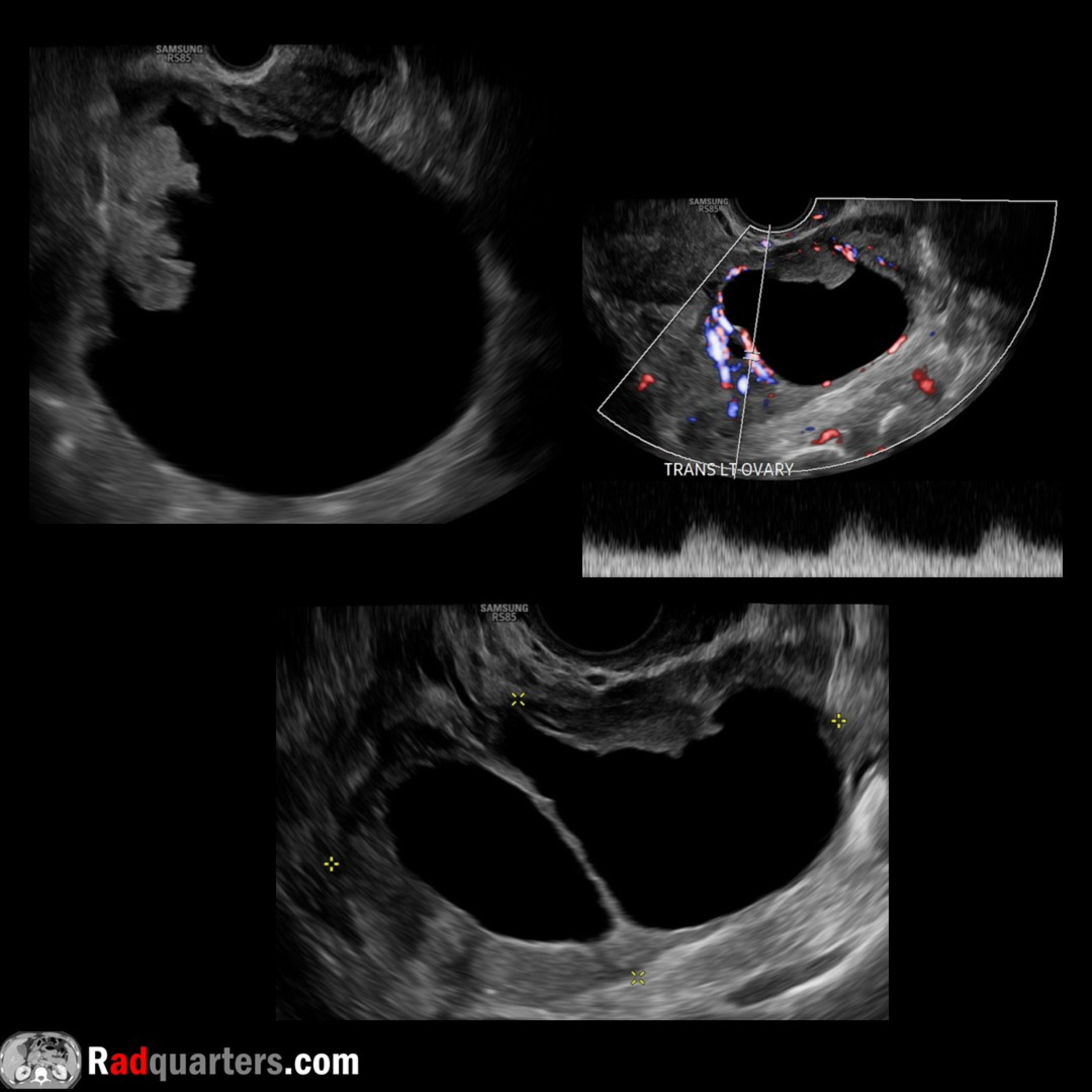 Radiology Lectures | RadquartersUltrasound of Ovarian Serous CystadenocarcinomaIn this radiology lecture, we review the ultrasound appearance of ovarian serous cystadenocarcinoma!
Key teaching points include:
Serous cystadenocarcinoma is the common ovarian malignancy and most common ovarian epithelial tumor
High-grade and low-grade types
Peak incidence 6th-7th decades
Ultrasound appearance: Mixed cystic and solid mass with papillary projections and thick septations
Elevated CA-125 in greater than 90%
Serous tumors are more commonly bilateral than other tumors
Four main categories of ovarian neoplasms: Epithelial (most common), germ cell (second most common), sex cord-stromal and metastases
Epithelial ovarian tumors are thought to originate outside the ovary (within fallopian...2024-05-0206 min
 Radiology Lectures | RadquartersUltrasound of Parathyroid AdenomaIn this radiology lecture, we review the ultrasound appearance of parathyroid adenoma!
Key teaching points include:
Benign tumor of the parathyroid glands
Most common cause of primary hyperparathyroidism: Elevated serum calcium and parathyroid hormone (PTH) levels
Ultrasound: Solid, homogeneous and very hypoechoic. Oval or bean-shaped, long axis oriented craniocaudal. Hypervascular. Majority posterior and inferior to thyroid. Hyperechoic line often separates adenoma from adjacent thyroid. Atypical features: Cystic degeneration, calcification.
Tc-99m sestamibi: Radiotracer uptake persisting on delayed 2-hour images. Taken up by both thyroid and parathyroid tissue, but washes out more rapidly from thyroid. Greater than 90...2024-04-0406 min
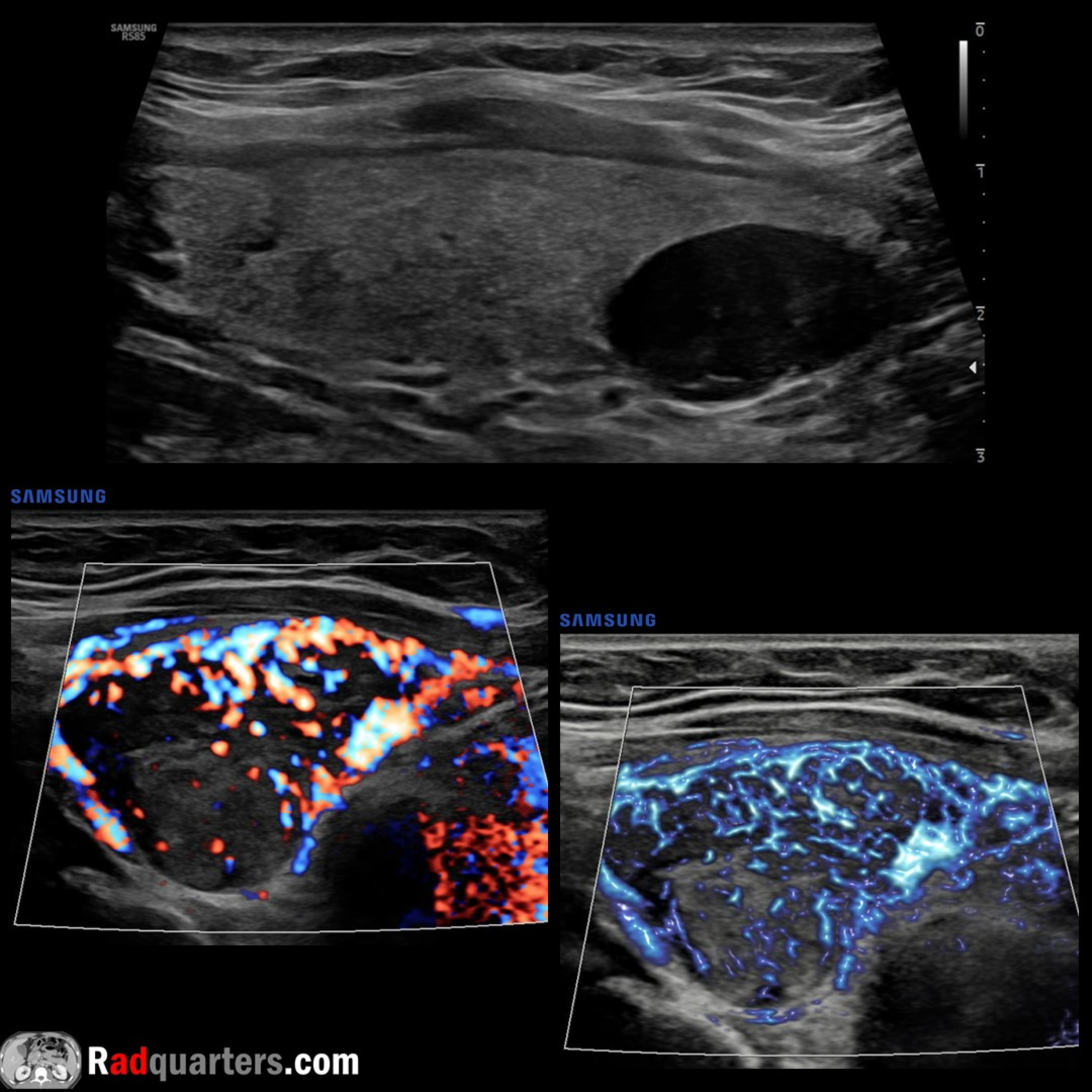
 Radiology Lectures | RadquartersUltrasound of ParotitisIn this radiology lecture, we review the ultrasound appearance of parotitis in the pediatric population!
Key teaching points include:
Parotitis = Inflammation of the parotid glands
Acute parotitis is usually infectious, most commonly viral
Mumps is most common viral cause in children, often bilateral
Bacterial parotitis can cause suppurative parotitis seen in premature infants and immunosuppressed children
Acute parotitis on US: Enlarged, heterogeneous, hyperemic gland(s) +/- lymphadenopathy
Since can be bilateral, comparison scanning essential
Bacterial parotitis may be complicated by abscess
“Pomegranate sign” may be seen in setting of acute parotitis: Uniform anechoic foci scattered throughout the...2024-03-0706 min
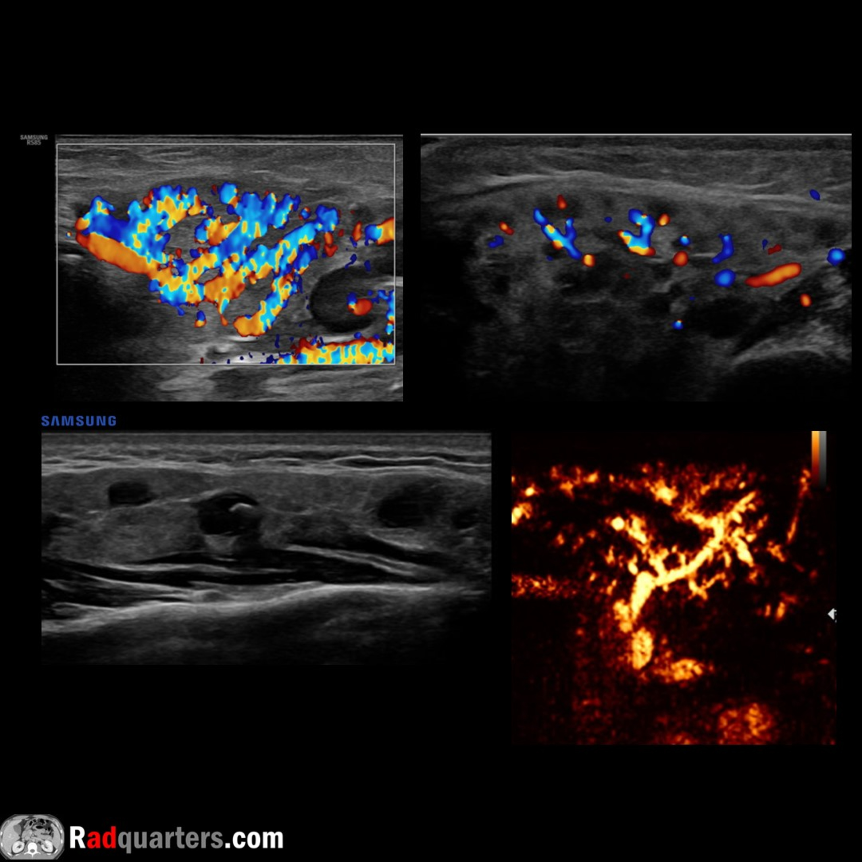
 Radiology Lectures | RadquartersUltrasound of Sublingual Dermoid CystIn this radiology lecture, we review the ultrasound appearance of sublingual dermoid cyst and explain floor of mouth anatomy!
Key teaching points include:
The floor of the mouth is a horseshoe-shaped area beneath tongue and in between sides of mandible, inferiorly bounded by mylohyoid muscle, and containing sublingual space (SLS)
SLS medial border: Midline genioglossus/geniohyoid muscle complex; SLS inferolateral border: Mylohyoid muscle
Anterior margin of hyoglossus muscle projects into posterior SLS
Sublingual dermoid cyst is a rare, benign cyst with squamous epithelial lining and contains skin appendages
Dermoid and epidermoid cysts are in same family...2024-02-0808 min
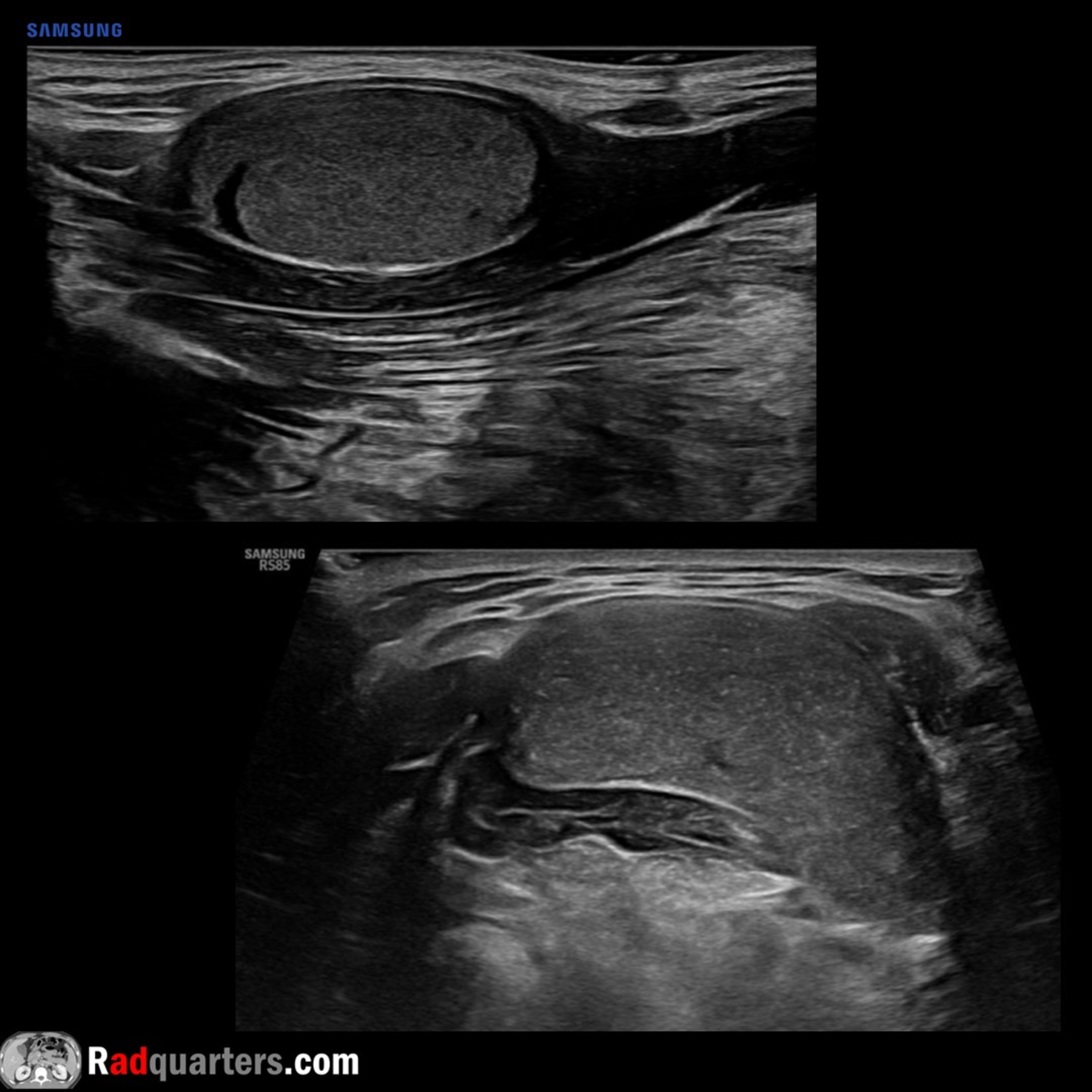
 Radiology Lectures | RadquartersUltrasound of Carpal Tunnel SyndromeIn this radiology lecture, we review the ultrasound appearance of carpal tunnel syndrome!
Key teaching points include:
Most common upper extremity entrapment neuropathy. Results from median nerve compression
With carpal tunnel syndrome, see hypoechoic enlargement of the median nerve as enters carpal tunnel with flattening of nerve = Notch sign, also volar bowing of flexor retinaculum
Median nerve area: Less than 8 mm2 = Normal; 8-12 mm2 = Borderline; greater than 12 mm2 = Abnormal
Most accurate to compare nerve area at proximal pronator quadratus muscle and carpal tunnel: Increase of 2 mm2 or more from proximal to distal = 99% sensitive and 100% specific for...2023-10-3009 min
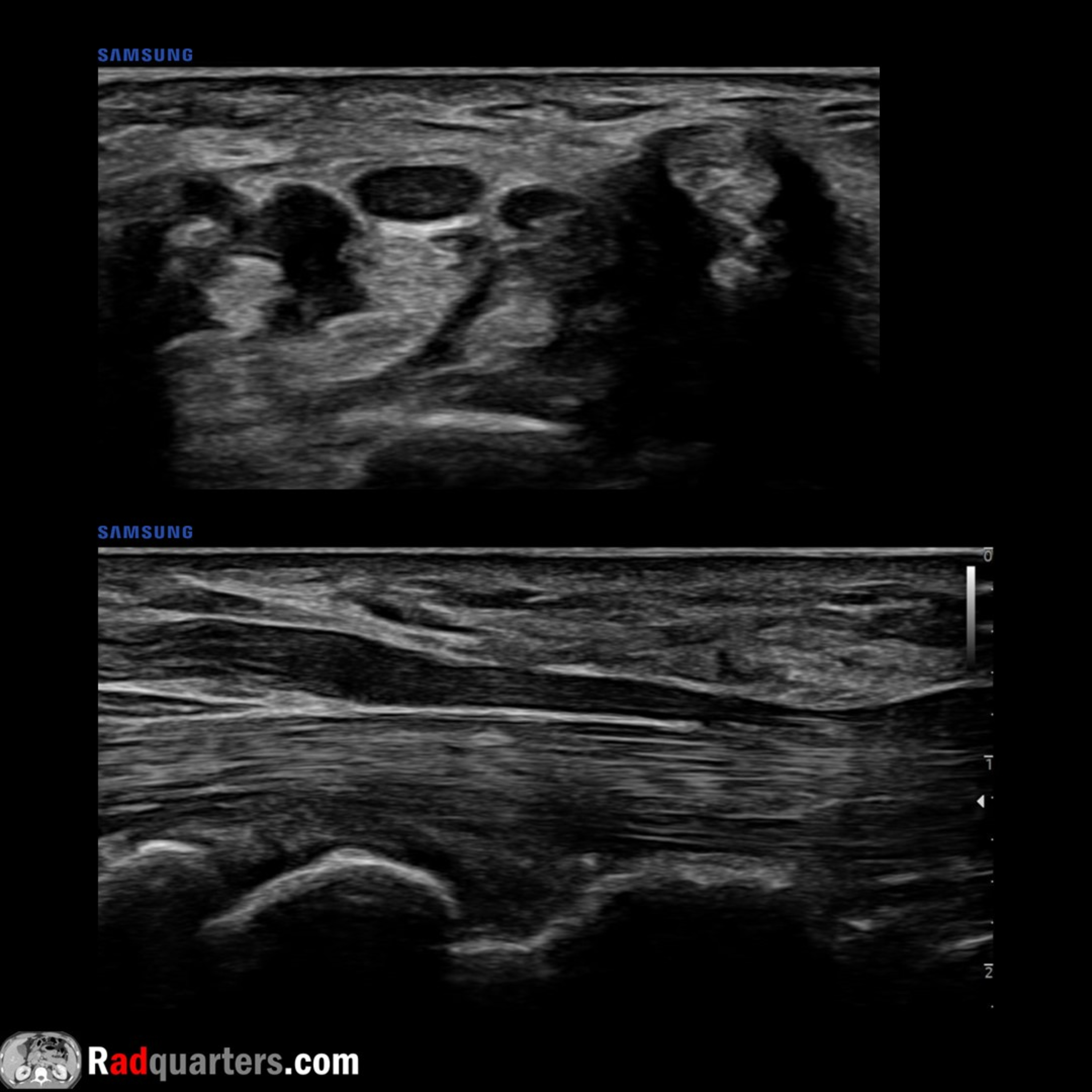
 Radiology Lectures | RadquartersUltrasound of Ganglion Cyst & Wrist Anatomy ReviewIn this radiology lecture, we review the ultrasound appearance of ganglion cysts while highlighting relevant wrist ultrasound anatomy!
Key teaching points include:
Ganglion cysts are viscous, mucin-filled collections lacking a synovial lining
Most commonly occur at hand/wrist = Most common wrist mass
Location: Dorsum of wrist (60%), frequently adjacent to scapholunate ligament; volar wrist (20%), often between radial artery and flexor carpi radialis tendon; flexor tendon sheath (10%); associated with DIP joint (10%)
Grows out of tissues surrounding joint like a balloon on a stalk. May see a pedicle connecting to joint
Usually well-defined and multilocular, can be unilocular
Hypoechoic...2023-09-2812 min
 Radiology Lectures | RadquartersRadquartersRadiologist Headquarters has a new name: Radquarters! Same high-yield content, but now with a streamlined name that’s easier to remember.
Click the YouTube Community tab or follow on social media for bonus teaching material posted throughout the week!
Spotify: https://spoti.fi/462r0F2
Instagram: https://www.instagram.com/Radquarters/
Facebook: https://www.facebook.com/Radquarters/
Twitter: https://twitter.com/Radquarters
Reddit: https://www.reddit.com/user/radiologistHQ/
The post Radquarters appeared first on Radquarters.
2023-09-2500 min
 Radiology Lectures | RadquartersRadquarters UpdateRadiologist Headquarters has a new name: Radquarters! Same high-yield content, but now with a streamlined name that's easier to remember.
Click the YouTube Community tab or follow on social media for bonus teaching material posted throughout the week!
Website: https://radquarters.com/
Instagram: https://www.instagram.com/radquarters/
Facebook: https://www.facebook.com/radquarters/
Twitter: https://twitter.com/radquarters
Reddit: https://www.reddit.com/user/radiologistHQ/
2023-09-2500 min
 Radiology Lectures | RadquartersUltrasound of Epididymitis & OrchitisIn this radiology lecture, we review the ultrasound appearance of acute epididymitis and orchitis!
Key teaching points include:
Epididymitis = Inflammation of epididymis. Usually bacterial, most commonly due to retrograde ascent from bladder or prostate.
Causative infectious agent varies based on age: Adults younger than 35: Neisseria gonorrhoeae, Chlamydia trachomatis (STDs). Adults older than 35: E. coli & other coliform bacteria.
Non-infectious causes of epididymitis: Trauma, repetitive activities such as sports (most common causes in males prior to sexual maturity), torsed appendix testis or appendix epididymis, vasculitis, and medications (amiodarone).
Presentation: Gradual onset of scrotal pain, swelling & urinary symptoms. Must...2023-08-2407 min
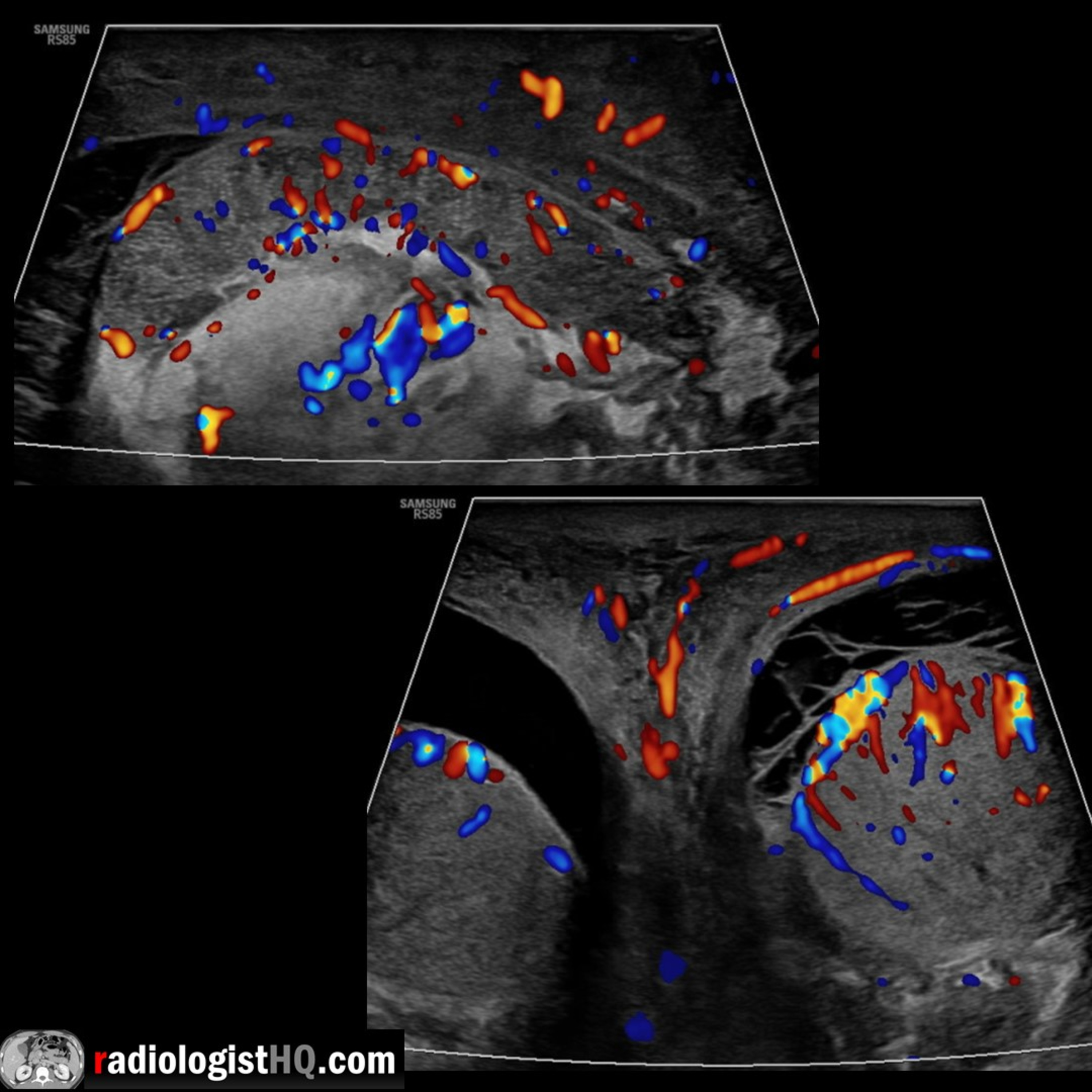
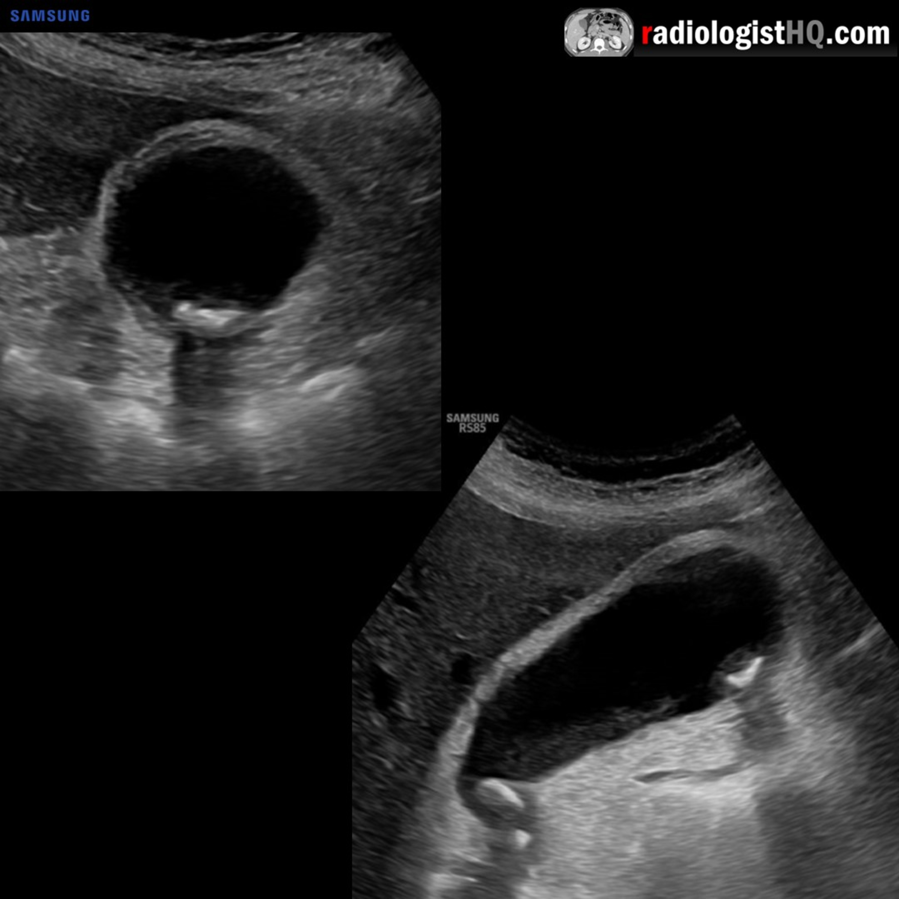
 Radiology Lectures | RadquartersUltrasound of Acute CholecystitisIn this radiology lecture, we review the ultrasound appearance of acute cholecystitis, including gangrenous and emphysematous cholecystitis!
Key teaching points include:
Acute cholecystitis = Acute gallbladder inflammation.
Most often (95%) caused by an impacted, obstructing gallstone in the cystic duct or gallbladder neck = Acute calculous cholecystitis.
Clinically presents as persistent RUQ pain that may radiate to right shoulder, often with N/V and fever.
Ultrasound findings of uncomplicated acute cholecystitis: Gallstones, sonographic Murphy sign, gallbladder wall thickening (greater than 3 mm) and edema, gallbladder distention (greater than 4 cm short axis), and pericholecystic fluid.
Sonographic Murphy sign = Maximal abdominal tenderness...2023-07-2710 min
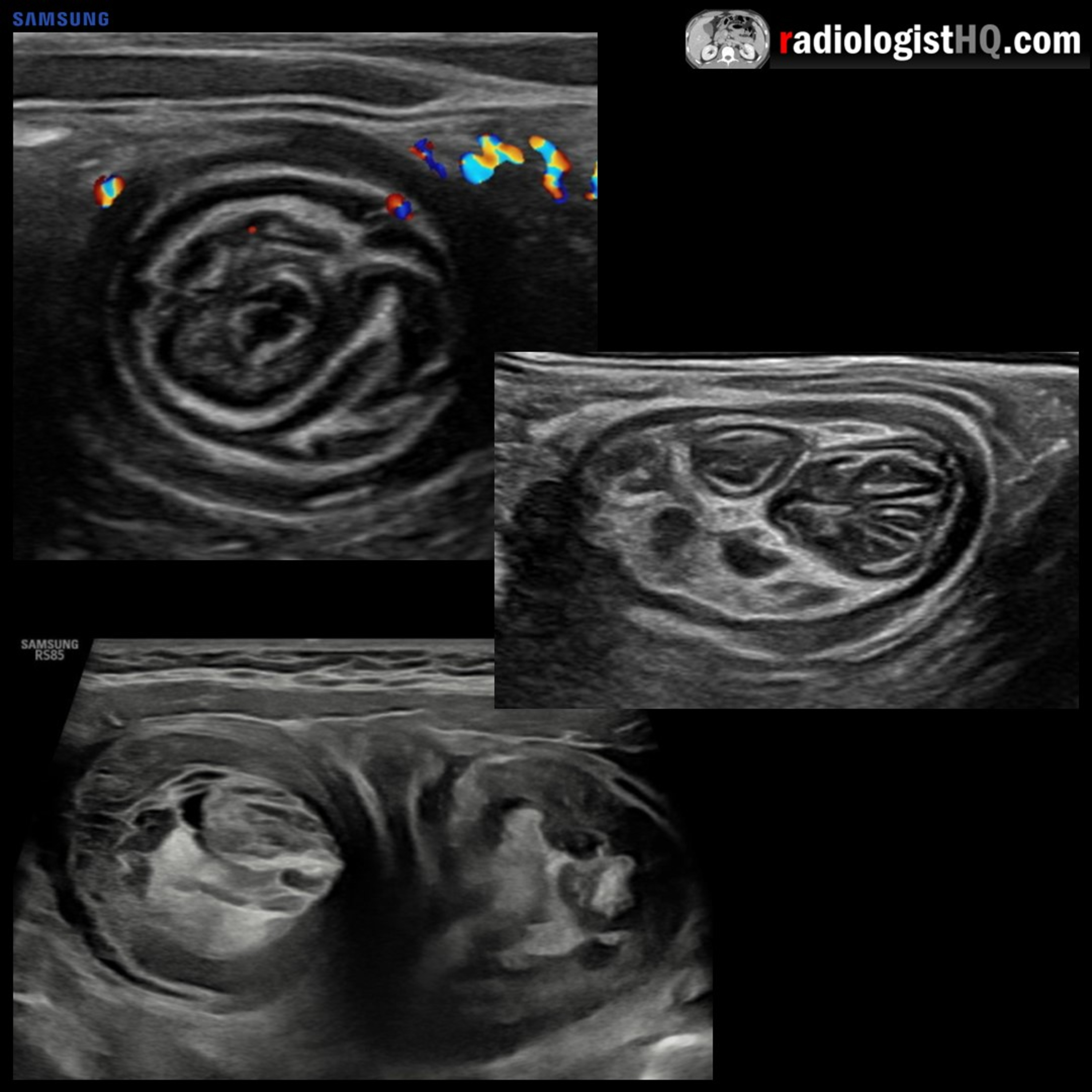 Radiology Lectures | RadquartersUltrasound of IntussusceptionIn this radiology lecture, we review the ultrasound appearance of ileocolic and small bowel-small bowel intussusception in children!
Key teaching points include:
Intussusception occurs when bowel is pulled into itself or into neighboring bowel.
Intussusceptum is the prolapsing bowel pulled into intussuscipiens which receives the bowel.
Two major types: Ileocolic and small bowel-small bowel.
If ileocolic not reduced = Bowel ischemia and perforation.
Most occur in children beyond 3 months of age.
Usually no lead point in children (unlike adults), suspected that due to hypertrophic lymphoid tissue after infection.
Clinical triad of colicky abdominal pain, vomiting, palpable abdominal...2023-05-2508 min
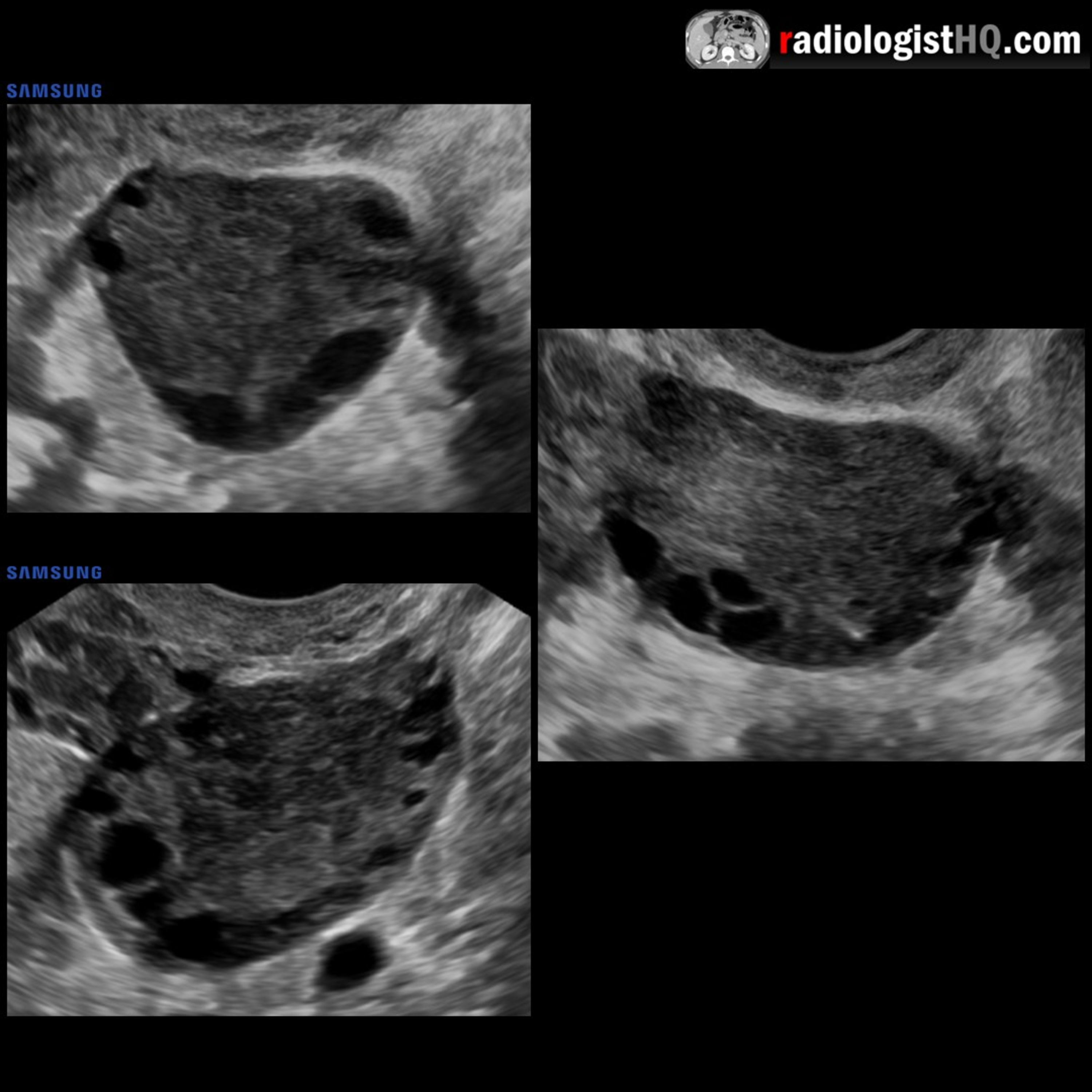
 Radiology Lectures | RadquartersUltrasound of Polycystic Ovarian SyndromeIn this radiology lecture, we review the ultrasound appearance of polycystic ovarian syndrome (PCOS)!
Key teaching points include:
PCOS often presents with the clinical triad of oligomenorrhea and/or anovulation, hirsutism, and obesity. Associated with subfertility and recurrent pregnancy loss.
Rotterdam criteria (2003) states that PCOS diagnosis requires at least two of the following: Oligo- or anovulation (ovulatory dysfunction), hyperandrogenism (clinical and/or biochemical signs), and polycystic ovarian morphology on ultrasound.
Ovaries can be sonographically normal in PCOS. “Hyperandrogenic anovulation” proposed as a more accurate term.
Ovaries can also appear polycystic on ultrasound without clinical diagnosis of PCOS...2023-04-2706 min
 Radiology Lectures | RadquartersUltrasound of Pleomorphic Adenoma of the Parotid GlandIn this radiology lecture, we review the ultrasound appearance of pleomorphic adenoma of the parotid gland!
Key teaching points include:
Pleomorphic adenoma AKA benign mixed tumor.
Most common salivary gland tumor, most common benign salivary gland tumor, and most common in the parotid gland.
Most common in patients aged 40-50, slightly more common in females.
For salivary gland masses in adults, the larger the gland, the more likely the tumor is benign: Parotid gland: 80%, submandibular gland: 50%, sublingual glands: 20%.
Parotid Gland 80% Rule: 80% of all salivary tumors are in the parotid, 80% of benign parotid gland tumors are pleomorphic...2023-03-0908 min
 Radiology Lectures | RadquartersUltrasound of Epidermal Inclusion CystIn this radiology lecture, we review the ultrasound appearance of epidermal inclusion cyst!
Key teaching points include:
Epidermal inclusion cyst is the most common cutaneous cyst.
Can occur anywhere: Head, neck, trunk, extremities.
Benign, keratin-containing cyst lined by a wall of stratified squamous epithelium.
On ultrasound, appears as a well-circumscribed, round to oval mass with broad (50%) contact with dermis, nonvascular and with posterior acoustic enhancement.
Hypoechoic to minimally hyperechoic with internal linear echogenic and anechoic debris = “Pseudotestis.”
Presence of a focal hypoechoic tract extending towards epidermis adds specificity = “Submarine sign.” May see overlying punctum on skin surface...2023-02-1608 min
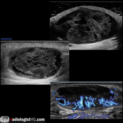
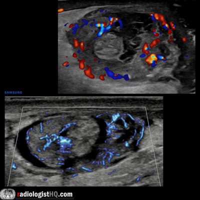 Radiology Lectures | RadquartersUltrasound of Torsion of the Appendix TestisIn this radiology lecture, we review the ultrasound appearance of torsion of the appendix testis and appendix epididymis!
Key teaching points include:
Appendix testis is a vestigial appendage usually located between upper pole of testis and head of epididymis.
AKA hydatid of Morgagni, the appendix testis is commonly present as a normal finding.
Appendix epididymis typically arises from epididymal head.
Both scrotal appendages are often pedunculated which increases risk of torsion.
Torsion occurs when appendage twists, occluding blood supply.
Torsion of the appendix testis is one of most common causes of acute scrotal pain in prepubertal...2023-01-1910 min
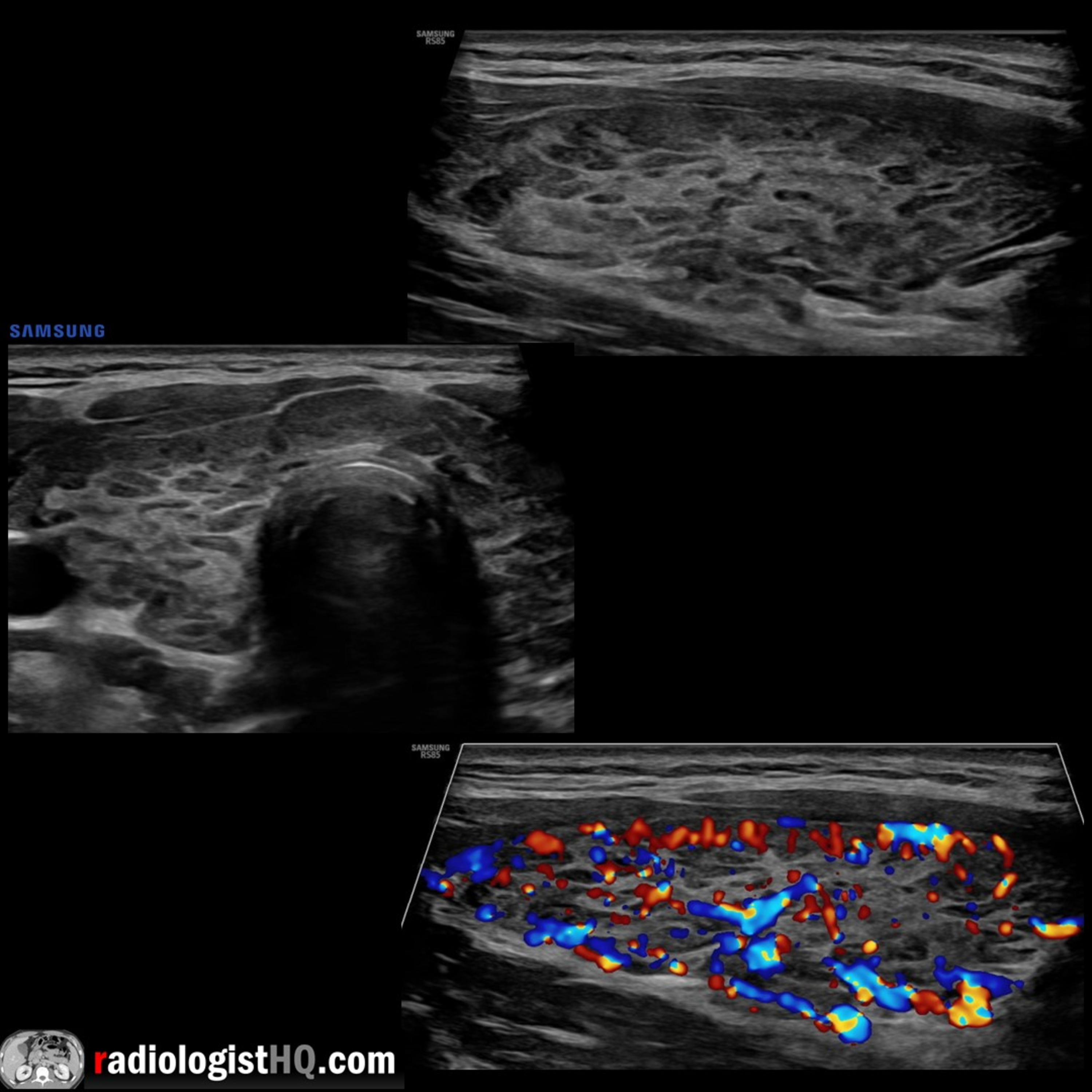 Radiology Lectures | RadquartersUltrasound of Hashimoto’s ThyroiditisIn this radiology lecture, we review the ultrasound appearance of Hashimoto’s thyroiditis with three unique cases!
Key teaching points include:
Normal thyroid gland isthmus measures less than 0.4 cm, transverse and AP lobe diameters measure less than 2 cm.
Hashimoto’s thyroiditis is an autoimmune thyroiditis caused by antibodies to thyroid proteins.
Most common in middle-aged females.
May coexist with other autoimmune disorders: Lupus, rheumatoid arthritis.
AKA chronic autoimmune lymphocytic thyroiditis: Gland is infiltrated with lymphocytes and plasma cells, fibrotic reaction replaces normal parenchyma.
Leads to hypothyroidism = Most common cause in USA.
Increased risk of thyroid cancer, inclu...2022-12-1508 min
 Radiology Lectures | RadquartersUltrasound of Hashimoto’s ThyroiditisIn this radiology lecture, we review the ultrasound appearance of Hashimoto’s thyroiditis with three unique cases!
Key teaching points include:
Normal thyroid gland isthmus measures less than 0.4 cm, transverse and AP lobe diameters measure less than 2 cm.
Hashimoto’s thyroiditis is an autoimmune thyroiditis caused by antibodies to thyroid proteins.
Most common in middle-aged females.
May coexist with other autoimmune disorders: Lupus, rheumatoid arthritis.
AKA chronic autoimmune lymphocytic thyroiditis: Gland is infiltrated with lymphocytes and plasma cells, fibrotic reaction replaces normal parenchyma.
Leads to hypothyroidism = Most common cause in USA.
Increased risk of thyroid cancer, incl...2022-12-1508 min
 Radiology Lectures | RadquartersUltrasound of Acute AppendicitisIn this radiology lecture, we review the ultrasound appearance of acute appendicitis with three unique cases!
Key teaching points include:
Ultrasound is the first-line imaging modality in pediatric and pregnant patients due to lack of ionizing radiation: Sensitivity/specificity approximately 80%.
Technique: Linear transducer with graded compression at site of maximal tenderness using gradual increased pressure to displace normal bowel gas.
Inflamed appendix appears as a noncompressible, blind-ending tubular structure arising from cecum.
Outer appendiceal diameter with compression: Less than 6 mm almost always normal, 6-8 mm borderline, greater than 8 mm highly suspicious.
Thickened appendiceal wall (greater than 2...2022-11-1712 min
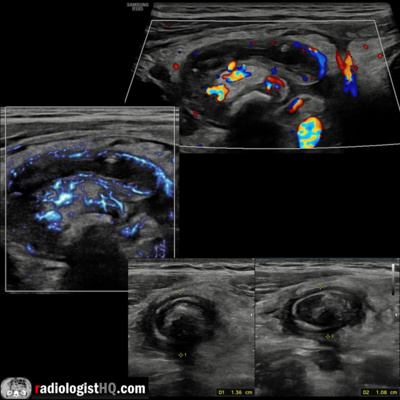
 Radiology Lectures | RadquartersUltrasound of Thyroglossal Duct CystIn this radiology lecture, we review the ultrasound appearance of thyroglossal duct cyst with two unique cases!
Key teaching points include:
Thyroglossal duct cyst is the most common congenital neck cyst.
Most present before age 18 as a midline, fluctuant neck mass near hyoid bone.
Often asymptomatic unless superinfected = Abscess, draining sinus.
Epithelial-lined cysts caused by failure of normal involution of thyroglossal duct.
Can occur anywhere from foramen cecum of tongue to thyroid gland.
Most are infrahyoid, followed by hyoid and suprahyoid.
Most are midline, but can be paramedian (more likely if infrahyoid).
If infrahyoid, typically embedded...2022-09-2908 min
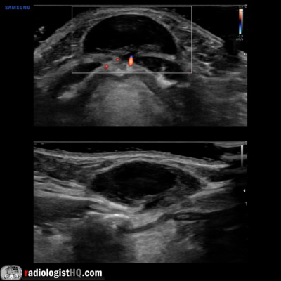 Radiology Lectures | RadquartersUltrasound of Thyroglossal Duct CystIn this radiology lecture, we review the ultrasound appearance of thyroglossal duct cyst with two unique cases
Key teaching points include:
Thyroglossal duct cyst is the most common congenital neck cyst.
Most present before age 18 as a midline, fluctuant neck mass near hyoid bone.
Often asymptomatic unless superinfected = Abscess, draining sinus.
Epithelial-lined cysts caused by failure of normal involution of thyroglossal duct.
Can occur anywhere from foramen cecum of tongue to thyroid gland.
Most are infrahyoid, followed by hyoid and suprahyoid.
Most are midline, but can be paramedian (more likely if infrahyoid).
If infrahyoid, typically embedded...2022-09-2908 min
 Radiology Lectures | RadquartersUltrasound of VaricoceleIn this radiology lecture, we review the ultrasound appearance of scrotal varicocele with three unique cases.
Key teaching points include:
Varicocele is abnormal dilatation of pampiniform venous plexus = Peritesticular veins.
Seen in up to 15% of adult and adolescent males.
Caused by incompetent or absent testicular vein valves.
Upper limit of normal for scrotal vein caliber = 2 mm, varicocele when greater than 2-3 mm.
Flow in varicocele usually too slow to detect with color Doppler and is typically better seen with Valsalva or with standing position.
85% left sided, 15% bilateral: Left testicular vein drains into left renal vein at 90...2022-08-3107 min
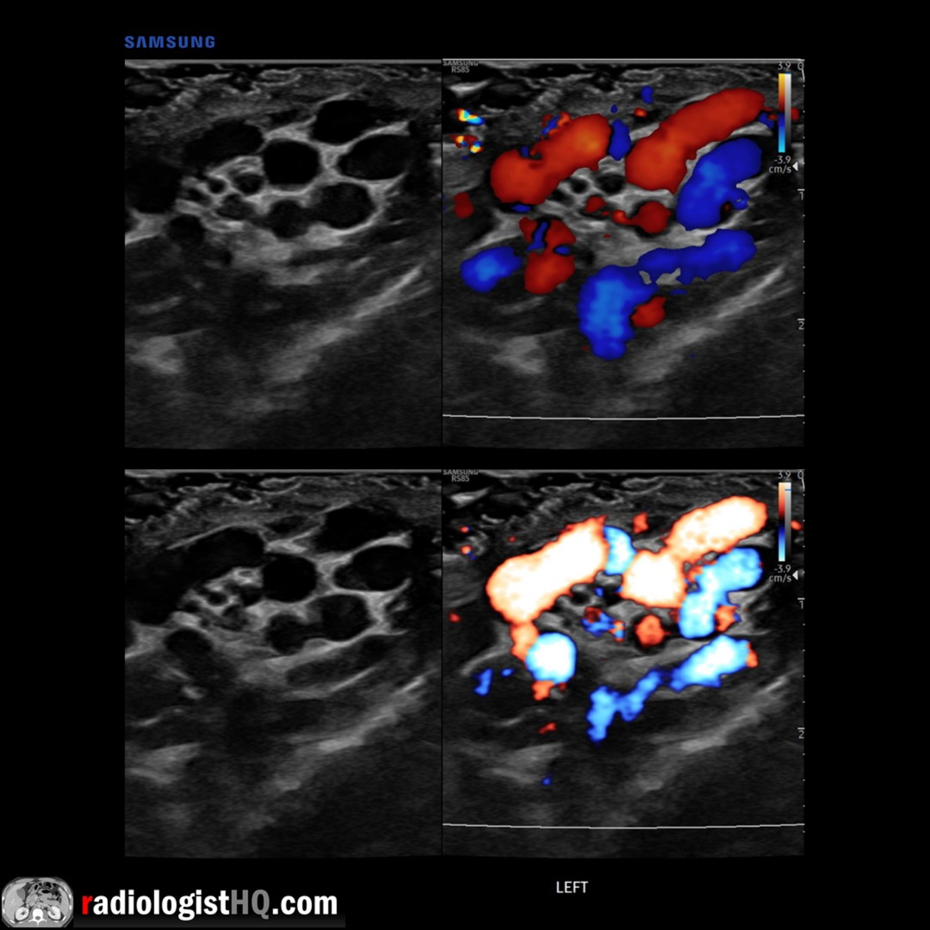
 Radiology Lectures | RadquartersUltrasound of Achilles Tendinosis and TearIn this radiology lecture, we review the ultrasound appearance of Achilles tendinosis, partial thickness tears and full thickness tears through four unique cases.
Key teaching points include:
Achilles tendon is strongest in body. Originates from soleus and gastrocnemius muscles, inserts onto posterior calcaneal tuberosity.
Achilles tendon tears = Most common ankle tendon injury.
Tendon enlargement greater than 1 cm in AP dimension = Abnormal.
Tendinosis appears as fusiform hypoechoic swelling of tendon without fiber disruption with increased blood flow (use power Doppler or microvascular flow).
Ultrasound highly sensitive and specific for partial and complete Achilles tears.
Partial tear = Hypoechoic...2022-07-2909 min
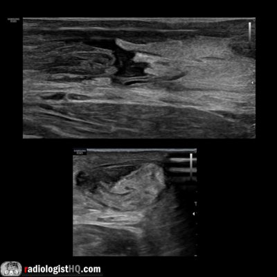
 Radiology Lectures | RadquartersUltrasound of Uterine AdenomyosisIn this radiology lecture, we review the ultrasound appearance of adenomyosis through three unique cases, including an MRI example.
Key teaching points include:
Adenomyosis results from ectopic endometrial tissue in myometrium. Leads to dysfunctional smooth muscle hyperplasia/hypertrophy surrounding ectopic glands.
Cause unknown.
Common, usually multiparous women of reproductive age.
Additional risk factors: Early menarche, short menstrual cycles, high BMI = High estrogen exposure.
Rarely seen in postmenopausal patients, unless treated with tamoxifen for breast cancer.
Often asymptomatic, but can present with menorrhagia, dysmenorrhea, dyspareunia, and chronic pelvic pain.
For diagnosing adenomyosis, transvaginal US much more sensitive...2022-06-2910 min
 Radiology Lectures | RadquartersUltrasound of EndometriomaIn this radiology lecture, we review the ultrasound appearance of endometrioma through three unique cases, including an MRI example.
Key teaching points include:
Endometriosis = Ectopic endometrial glands and stroma outside of the uterine cavity. Includes endometriomas, extraovarian implants and adhesions.
Endometriomas = Endometriotic cysts within ovary.
Endometriosis is seen in about 10% of women of reproductive age.
Presentation: Pelvic pain, dysmenorrhea, dyspareunia, infertility.
Ultrasound: Diffuse, homogeneous low-level echoes (most specific feature) yielding a ground glass appearance. May have posterior acoustic enhancement.
Endometriomas may have peripheral punctate echogenic foci. These foci have no internal vascular flow but can see...2022-05-2508 min
 Radiology Lectures | RadquartersUltrasound of Testicular TorsionIn this radiology lecture, we review the ultrasound appearance of testicular torsion through three unique cases.
Key teaching points include:
Torsion occurs when spermatic cord twists and cuts off blood supply to the testis.
Bell-clapper deformity most common etiology: Abnormally high attachment of tunica vaginalis allowing spermatic cord rotation and testicular torsion (intravaginal).
Torsion has a bimodal distribution: First year of life (extravaginal), adolescents/young adults (intravaginal).
“Whirlpool” sign: Eddy swirl of coiled spermatic cord superior to testis, highly specific but less commonly seen than redundant spermatic cord.
Redundant spermatic cord AKA boggy pseudomass, torsion knot, epid...2022-04-1309 min
 Radiology Lectures | RadquartersUltrasound & CT of Renal OncocytomaIn this radiology lecture, the ultrasound and CT appearance of renal oncocytoma is revealed.
Key teaching points include:
Oncocytomas are benign, solid tumors.
13% patients have multiple oncocytomas, and 1/3rd have concurrent renal cell carcinoma.
Central stellate nonenhancing scar only seen in 1/3rd of cases, and more commonly in larger tumors.
Spoke-wheel angiographic pattern may be present, best visualized on ultrasound using microvascular flow, AKA superb microvascular imaging.
Often not possible to differentiate from renal cell carcinoma (RCC) with imaging.
Both oncocytoma and RCC can have central scar and/or spoke-wheel angiographic pattern.
Kim et al.* found...2022-03-2306 min
 Radiology Lectures | RadquartersCase of the Week: Ovarian Mucinous Cystadenocarcinoma (Ultrasound & MRI)In this radiology lecture, we reveal the imaging appearance of mucinous cystadenocarcinoma of the ovary and explain differentiating features from serous cystadenocarcinoma.
Key points include:
A rare type of malignant ovarian epithelial tumor.
Often large at presentation, may be enormous.
Almost always multilocular.
Mucinous, proteinaceous and hemorrhagic material within loculi.
US: Scattered low-level echoes.
MRI: “Stained glass” appearance = Variable T1/T2 signal. Thick mucin = T1/T2 hyperintense.
Irregular, thick septations and solid components with internal vascularity and enhancement allow differentiation from mucinous cystadenoma.
Click the YouTube Community tab or follow on social media for bonus teac...2022-03-0208 min
 Radiology Lectures | RadquartersCase of the Week: Wandering Spleen (CT)In this radiology lecture, we discuss the CT appearance of wandering spleen!
Key points include:
Extremely rare, usually between 20-40 years of age, more common in females.
Splenic mobility due to congenital or acquired abnormality of the normal peritoneal attachments/suspensory ligaments.
Splenic migration to lower abdomen/pelvis, may develop long vascular pedicle.
Twisting of pedicle can lead to splenic ischemia and infarction if not promptly treated.
Variable clinical presentation, patients often become symptomatic if torsion of pedicle occurs: Intermittent colicky pain, vague abdominal discomfort, abdominal mass, acute abdomen.
Treatment: Surgical detorsion and fixation of spleen...2022-02-1603 min
 Radiology Lectures | RadquartersUltrasound of Complete Molar PregnancyIn this radiology lecture, the ultrasound appearance of complete molar pregnancy is revealed.
Key points include:
AKA hydatiform mole = Most common form of gestational trophoblastic disease.
Gestational trophoblastic neoplasia (GTN) less common = Invasive mole and choriocarcinoma.
Approximately 1/1,000 pregnancies is a molar pregnancy.
Most common in females under age 20 and over age 35.
Two types of molar pregnancy: Complete (most common) and partial.
Complete: Diploid (paternal DNA only), no fetus, more likely to be complicated by GTN.
Partial: Triploid (maternal and paternal DNA), abnormal fetus or fetal parts, harder to diagnose.
Complete hydatiform mole presentation: Vaginal bleeding, enlarged...2022-02-0905 min
 Radiology Lectures | Radquarters5 Cases in 5 Minutes: Musculoskeletal #4Quiz yourself with this week’s interactive video lecture as we present a total of 5 interesting musculoskeletal radiology cases followed by a diagnosis reveal and key teaching points after each case, all in just a few minutes!
Click the YouTube Community tab or follow on social media for bonus teaching material posted throughout the week!
Instagram: https://www.instagram.com/radiologistHQ/
Facebook: https://www.facebook.com/radiologistHeadQuarters/
Twitter: https://twitter.com/radiologistHQ
The post 5 Cases in 5 Minutes: Musculoskeletal #4 appeared first on Radquarters.
2022-02-0209 min
 Radiology Lectures | RadquartersCase of the Week: Septate Uterus (MRI)In this radiology lecture, we discuss the MRI appearance of septate uterus, and explain how to differentiate from other uterine anomalies.
Key points include:
Most common müllerian duct anomaly (55%): Septal reabsorption abnormality.
Ultrasound and MRI provide assessment of external uterine contour and presence of renal anomalies.
Hysterosalpingogram of limited value, cannot reliably differentiate between subtypes.
On MRI, uterine fundus is typically convex or minimally indented: Fundal cleft less than 1 cm.
Midline septum of variable length, may be muscular or fibrous.
Important to differentiate type of septum as may alter surgical approach.
Compared to bicornuate uterus, h...2022-01-2606 min
 Radiology Lectures | RadquartersUltrasound of Ovarian Dermoid CystIn this radiology lecture, we discuss the ultrasound appearance of ovarian dermoid cyst, including the rarely seen but highly specific “floating sphere” sign!
Key points include
AKA mature cystic teratoma.
Most common ovarian neoplasm.
Benign, mean age 30.
10% bilateral.
Mature tissue from ≥2 embryonic germ cell layers: Sebaceous material, hair follicles, skin derivatives, fat, muscle, bone, and other tissues lined by squamous epithelium.
Specificity of US diagnosis 94-100%.
MRI for changing morphology on f/u and for postmenopausal patients.
Ultrasound findings: Floating echogenic spherical structures = “Floating sphere” sign (uncommon but pathognomonic), hyperechoic component with acoustic shadowing (Rokitanksy nodule), hyperechoi...2022-01-1905 min
 Radiology Lectures | RadquartersCase of the Week: Retroperitoneal Fibrosis (Ultrasound & CT)In this radiology lecture, we discuss the ultrasound and CT appearance of retroperitoneal fibrosis.
Key points include:
Most cases (70%) are idiopathic = Ormond disease.
Nonspecific symptoms depending on involved structures: Malaise, weight loss, low-grade fever.
Ureteral entrapment: Obstructive uropathy or renal failure, may see medial deviation of middle third of ureters with hydronephrosis.
Venous entrapment: Lower extremity edema, deep venous thrombosis.
CT: Soft tissue mass anterolateral to aorta with posterior sparing.
DDx: Retroperitoneal Lymphoma will Lift the aorta.
MRI: Low T1/T2 signal when inactive, T2 bright with early enhancement when active inflammation.
PET/CT: Avid when...2022-01-1204 min
 Radiology Lectures | RadquartersCase of the Week: Perihilar Cholangiocarcinoma/Klatskin Tumor (CT & MRI)In this radiology lecture, we discuss the CT and MRI appearance of perihilar cholangiocarcinoma.
Key points include:
Perihilar cholangiocarcinoma (AKA Klatskin tumor) occurs at bifurcation of the hepatic duct.
Cholangiocarcinoma (CC) is a primary malignant tumor of bile duct epithelium, usually adenocarcinoma.
CC is the most common primary hepatic malignancy after hepatocellular carcinoma (HCC), and most are extrahepatic (as opposed to intrahepatic).
Appearance of CC is based on growth pattern: Mass-forming, periductal infiltrating, and intraductal growing.
Risk factors: Parasite infection, choledochal cyst, primary sclerosing cholangitis, recurrent pyogenic cholangitis, and inflammatory bowel disease (ulcerative colitis).
Patients are...2022-01-0509 min
 Radiology Lectures | RadquartersCase of the Week: Medullary Sponge Kidney (Ultrasound & CT)Join me in this radiology lecture revealing the ultrasound and CT appearance of medullary sponge kidney (MSK).
Key points include:
MSK is a developmental ectasia with cystic dilatation of the collecting tubules in the pyramids leading to medullary nephrocalcinosis.
DDx medullary nephrocalcinosis: Hyperparathyroidism (most common cause in adults), renal tubular acidosis (type 1), MSK, hypervitaminosis D, other causes of hypercalcemia, sarcoidosis.
MSK associations: Beckwith-Wiedemann syndrome, congenital hemihypertrophy, Caroli disease, Ehlers-Danlos syndrome.
US: Echogenic medullary pyramids.
CT: Renal calculi, striated nephrogram, excretory phase “paintbrush” appearance or “growing calculus” sign.
Often asymptomatic but may present due to renal stones.
Click...2021-12-2206 min
 Radiology Lectures | RadquartersCase of the Week: Amebic Liver Abscess (Ultrasound & CT)In this radiology lecture, we discuss the ultrasound and CT appearance of amebic liver abscess.
Key points include:
Entamoeba histolytica infection.
Endemic in Africa, Southeast Asia, and Central & South America.
More common in males.
Presents as right upper quadrant pain, fever and hepatomegaly.
Both amebic and pyogenic (bacterial) abscesses can have a layered wall with the “double target” or “double rim” sign.
Amebic more likely to be unilocular (septations present in 30%) without “cluster” sign typical of multiloculated pyogenic abscess.
Amebic more likely solitary, pyogenic more likely multiple.
Can be treated medically (metronidazole), but if diagnosis uncertain, if there is...2021-12-1505 min
 Radiology Lectures | RadquartersCase of the Week: Pulmonary Infarction (X-ray & CT)In this radiology lecture, we discuss the chest x-ray and CT appearance of pulmonary infarction in the setting of acute pulmonary embolism.
Key points include:
Uncommon complication of pulmonary embolism.
Most common in right lung.
Risk of infarction increases with large clot burden.
Typically wedge-shaped, peripheral consolidation with no air bronchograms (Hampton hump).
However, may not be wedge-shaped, and not all wedge-shaped opacities will be infarcts in the setting of pulmonary embolism.
“Bubbly” consolidation containing rounded, central lucencies: Most specific finding of infarct* and represents a combination of infarcted, necrotic lung and adjacent viable, aerated lung.
“Vessel...2021-12-0805 min
 Radiology Lectures | RadquartersCase of the Week: Ruptured Ectopic Pregnancy (Ultrasound)In this radiology lecture, we discuss the ultrasound appearance of ruptured ectopic pregnancy.
Key points include:
Most ectopic pregnancies occur in the fallopian tube: Ampulla most common, followed by isthmus and fimbria.
Risk factors: Prior ectopic pregnancy, prior surgery (fallopian tube), pelvic inflammatory disease, endometriosis, IVF.
“A single measurement of hCG, regardless of its level, does not reliably distinguish between ectopic and intrauterine pregnancy (viable or nonviable).”*
Levels of hCG in ectopic pregnancies are highly variable.
Tubal rupture main complication, occurs in up to 20%.
Free fluid in pelvis alone nonspecific, but echogenic fluid in Morison pouch (subh...2021-12-0105 min
 Radiology Lectures | RadquartersCase of the Week: Gallstone Ileus (X-ray & CT)In this radiology lecture, we discuss the appearance of gallstone ileus on x-ray and CT.
Key points include:
Gallstone ileus is a rare complication of chronic cholecystitis.
Actually not an ileus, but a small bowel obstruction.
Gallstone migrates through a fistula between gallbladder and small bowel (usually duodenum) and becomes impacted in the terminal ileum.
Stone can also impact in the proximal ileum, jejunum, even in the duodenum/distal stomach causing gastric outlet obstruction (Bouveret syndrome).
Rigler triad on abdominal x-ray: Small bowel obstruction, pneumobilia and gallstone in the right iliac fossa.
Usually affects the elderly...2021-11-2404 min
 Radiology Lectures | RadquartersCase of the Week: Testicular Epidermoid Cyst (Ultrasound)In this radiology lecture, we discuss the ultrasound appearance of testicular epidermoid cyst.
Key points include:
Testicular epidermoid cyst is a rare, benign, intratesticular neoplasm.
Most common in 2nd-4th decades, typically presents as a painless mass.
Lamellated, onion-like, bull’s-eye appearance: Alternating hyperechoic and hypoechoic concentric rings.
Appearance secondary to cyst filled with layers of keratin and lined with keratinizing squamous epithelium.
Non-vascular and sharply marginated.
Nonenhancing on MRI.
Important to recognize preoperatively because may be treated with conservative surgery.
Management somewhat controversial as originally diagnosed with orchiectomy.
Increasingly treated with enucleation if frozen sections of...2021-11-1702 min
 Radiology Lectures | RadquartersCase of the Week: Necrotizing Pancreatitis (CT & MRI)In this radiology lecture, we discuss the imaging appearance of necrotizing pancreatitis on both CT and MRI.
Key points include:
According to the revised Atlanta classification, there are two types of acute pancreatitis: Interstitial edematous pancreatitis (IEP) and necrotizing pancreatitis (NP).
For IEP, fluid collection in first 4 weeks = acute peripancreatic fluid collection, after 4 weeks = pseudocyst.
For NP, fluid collection in first 4 weeks = acute necrotic collection, after 4 weeks = walled-off necrosis.
Non-enhancing hypoattenuating areas = necrotizing pancreatitis.
Gas suspicious for infection/emphysematous pancreatitis.
Vascular complications are important to identify.
Venous thrombosis: splenic, portal, and mesenteric veins.
Pseudoaneurysms: Splenic and...2021-11-1007 min
 Radiology Lectures | RadquartersCase of the Week: Duplicated Collecting System (VCUG & Ultrasound)In this radiology lecture, we discuss the imaging appearance of duplicated collecting system and ureterocele, with attention to US and VCUG.
Key points include:
Weigert-Meyer rule: Remember the mnemonic “DUMI.”
With duplex kidneys and complete ureteral Duplication, ureter draining Upper pole inserts ectopically into bladder Medially and Inferiorly to ureter draining lower pole.
Lower pole moiety inserts orthotopically.
Upper pole moiety often ends as an ectopic ureterocele.
Upper pole moiety tends to obstruct, and lower pole moiety is prone to reflux.
Obstructed upper pole moiety causes mass effect with resultant inferior displacement of the lower pole moie...2021-11-0305 min
 Radiology Lectures | RadquartersCase of the Week: Colonic Lymphoma (CT & PET)In this radiology lecture, we discuss the imaging appearance of large bowel lymphoma.
Key points include:
Often isodense to skeletal muscle.
May have aneurysmal dilatation of involved bowel.
Less likely obstructive and longer segment involvement compared to colonic adenocarcinoma.
Located near ileocecal valve.
GI lymphoma: Most common in stomach, followed by small bowel (ileum, jejunum, duodenum), least common site colorectal.
Splenomegaly and severe lymphadenopathy favor lymphoma but may not be present.
Click the YouTube Community tab or follow on social media for bonus teaching material posted throughout the week!
Instagram: https://www.instagram...2021-10-2703 min
 Radiology Lectures | RadquartersCase of the Week: Ovarian Torsion (Ultrasound)In this radiology lecture, we discuss the ultrasound appearance of ovarian torsion.
Key points include:
Rotation of ovarian vascular pedicle with obstruction to venous outflow and arterial inflow.
Enlarged, heterogenous ovary due to hemorrhage and edema.
Small peripheral cysts with “follicular ring” sign: Thick, hyperechoic rim surrounding follicles of torsed ovary.
Classically, absent vascular flow.
However, up to 60% of patients with torsion have normal color Doppler findings, and arterial flow may even be preserved!
Most common finding is decreased or absent venous flow.
Twisted vascular pedicle: Most definitive sign of torsion, present in up to 88% of pati...2021-10-2003 min
 Radiology Lectures | RadquartersCase of the Week: Cecal Volvulus (X-ray & CT)In this inaugural case of the week radiology lecture, we discuss the imaging appearance of cecal volvulus!
Key points include:
Cecal volvulus occurs when there is twisting of cecum around the mesentery with proximal large bowel obstruction.
Cecum normally 2021-10-1305 min
 Radiology Lectures | RadquartersWebinar: Ultrasound of Ovarian Cystic DiseaseCheck out my FREE webinar titled “Ultrasound of Ovarian Cystic Disease” on Wednesday, September 29, 2021 at 7-8 PM ET, hosted by the American Institute of Ultrasound in Medicine (AIUM). A Q&A session follows the presentation.
In case you missed the lecture or would like to watch it again, click here: https://learn.aium.org/products/ultrasound-of-cystic-ovarian-disease-neoplastic-non-neoplastic
Enjoy!
The post Webinar: Ultrasound of Ovarian Cystic Disease appeared first on Radquarters.
2021-09-2700 min
 The Psychology PodcastMarcus Kowal - Healing Through MeaningThe 24th episode of the Psychology Podcast with Daniel Karim features Marcus Kowal, fighter, entrepreneur, activist, and author.
Marcus is a decorated MMA fighter, Brazilian Jiu Jitsu competitor, kickboxing black belt, and a second-degree Krav Maga black belt. He is also an author, accomplished business owner, entrepreneur, trainer, life coach, and activist.
He’s the founder and owner of Systems Training Center, with four Los Angeles-area locations, and has expanded his reach online with the platforms YourKravMaga.com and the NEOU Fitness App. Now, most of his time is spent on the other side of the mat as an instructor an...2021-07-3043 min
 Radiology Lectures | RadquartersWebinar: Ultrasound of Genitourinary Infectious DiseaseCheck out my FREE webinar titled “Ultrasound of Genitourinary Infectious Disease” on Thursday, March 25, 2021 at 1-2 PM ET, hosted by the American Institute of Ultrasound in Medicine (AIUM). A Q&A session follows the presentation.
In case you missed the lecture or would like to watch it again, you can find it here: https://learn.aium.org/products/ultrasound-of-genitourinary-infectious-disease
Enjoy!
The post Webinar: Ultrasound of Genitourinary Infectious Disease appeared first on Radquarters.
2021-03-2200 min
 Radiology Lectures | Radquarters5 Cases in 5 Minutes: Head & Neck #2Quiz yourself with this week’s interactive video lecture as we present a total of 5 interesting head and neck radiology cases followed by a diagnosis reveal and key teaching points after each case, all in just a few minutes!
The post 5 Cases in 5 Minutes: Head & Neck #2 appeared first on Radquarters.
2019-07-0411 min
 Radiology Lectures | Radquarters5 Cases in 5 Minutes: Musculoskeletal #3Quiz yourself with this week’s interactive video lecture as we present a total of 5 interesting musculoskeletal radiology cases followed by a diagnosis reveal and key teaching points after each case, all in just a few minutes!
The post 5 Cases in 5 Minutes: Musculoskeletal #3 appeared first on Radquarters.
2019-06-2710 min
 Radiology Lectures | Radquarters5 Cases in 5 Minutes: Musculoskeletal #2Quiz yourself with this week’s interactive video lecture as we present a total of 5 interesting musculoskeletal radiology cases followed by a diagnosis reveal and key teaching points after each case, all in just a few minutes!
The post 5 Cases in 5 Minutes: Musculoskeletal #2 appeared first on Radquarters.
2019-06-2011 min
 Radiology Lectures | Radquarters5 Cases in 5 Minutes: Head & Neck #1Quiz yourself with this week’s interactive video lecture as we present a total of 5 interesting head and neck imaging cases followed by a diagnosis reveal and key teaching points after each case, all in just a few minutes!
The post 5 Cases in 5 Minutes: Head & Neck #1 appeared first on Radquarters.
2019-06-1312 min
 Radiology Lectures | Radquarters5 Cases in 5 Minutes: Musculoskeletal #1Quiz yourself with this week’s interactive video lecture as we present a total of 5 interesting musculoskeletal imaging cases followed by a diagnosis reveal and key teaching points after each case, all in just a few minutes!
The post 5 Cases in 5 Minutes: Musculoskeletal #1 appeared first on Radquarters.
2019-06-0609 min
 Radiology Lectures | Radquarters5 Cases in 5 Minutes: Vascular #5Quiz yourself with this week’s interactive video lecture as we present a total of 5 interesting vascular imaging cases followed by a diagnosis reveal and key teaching points after each case, all in just a few minutes!
The post 5 Cases in 5 Minutes: Vascular #5 appeared first on Radquarters.
2019-05-3009 min
 Radiology Lectures | Radquarters5 Cases in 5 Minutes: Thoracic #5Quiz yourself with this week’s interactive video lecture as we present a total of 5 interesting thoracic imaging cases followed by a diagnosis reveal and key teaching points after each case, all in just a few minutes!
The post 5 Cases in 5 Minutes: Thoracic #5 appeared first on Radquarters.
2019-05-2310 min
 Radiology Lectures | Radquarters5 Cases in 5 Minutes: Vascular #4Quiz yourself with this week’s interactive video lecture as we present a total of 5 interesting vascular imaging cases followed by a diagnosis reveal and key teaching points after each case, all in just a few minutes!
The post 5 Cases in 5 Minutes: Vascular #4 appeared first on Radquarters.
2019-05-1611 min
 Radiology Lectures | Radquarters5 Cases in 5 Minutes: Thoracic #4Quiz yourself with this week’s interactive video lecture as we present a total of 5 unknown thoracic imaging cases followed by a diagnosis reveal and key teaching points after each case, all in just a few minutes!
The post 5 Cases in 5 Minutes: Thoracic #4 appeared first on Radquarters.
2019-05-0911 min
 Radiology Lectures | Radquarters5 Cases in 5 Minutes: Vascular #3Quiz yourself with this week’s interactive video lecture as we present a total of 5 unknown vascular imaging cases followed by a diagnosis reveal and key teaching points after each case, all in just a few minutes!
The post 5 Cases in 5 Minutes: Vascular #3 appeared first on Radquarters.
2019-05-0211 min
 Radiology Lectures | Radquarters5 Cases in 5 Minutes: Vascular #2Quiz yourself with this week’s interactive video lecture as we present a total of 5 unknown vascular imaging cases followed by a diagnosis reveal and key teaching points after each case, all in about 5(ish) minutes!
The post 5 Cases in 5 Minutes: Vascular #2 appeared first on Radquarters.
2019-04-2512 min
 Radiology Lectures | Radquarters5 Cases in 5 Minutes: Thoracic #3Quiz yourself with this week’s interactive video lecture as we present a total of 5 unknown thoracic imaging cases followed by a diagnosis reveal and key teaching points after each case, all in about 5(ish) minutes!
The post 5 Cases in 5 Minutes: Thoracic #3 appeared first on Radquarters.
2019-04-1809 min
 Radiology Lectures | Radquarters5 Cases in 5 Minutes: Thoracic #2Quiz yourself with this week’s interactive video lecture as we present a total of 5 unknown thoracic imaging cases followed by a diagnosis reveal and key teaching points after each case, all in about 5(ish) minutes!
The post 5 Cases in 5 Minutes: Thoracic #2 appeared first on Radquarters.
2019-04-1108 min
 Radiology Lectures | Radquarters5 Cases in 5 Minutes: Vascular #1Join us in this interactive video lecture as we present a total of 5 unknown vascular imaging cases followed by a diagnosis reveal and key teaching points after each case, all in about 5(ish) minutes!
The post 5 Cases in 5 Minutes: Vascular #1 appeared first on Radquarters.
2019-04-0410 min
 Radiology Lectures | Radquarters5 Cases in 5 Minutes: Thoracic #1Join us in this interactive video lecture as we present a total of 5 unknown thoracic imaging cases followed by a diagnosis reveal and key teaching points after each case, all in about 5(ish) minutes!
The post 5 Cases in 5 Minutes: Thoracic #1 appeared first on Radquarters.
2019-03-2809 min
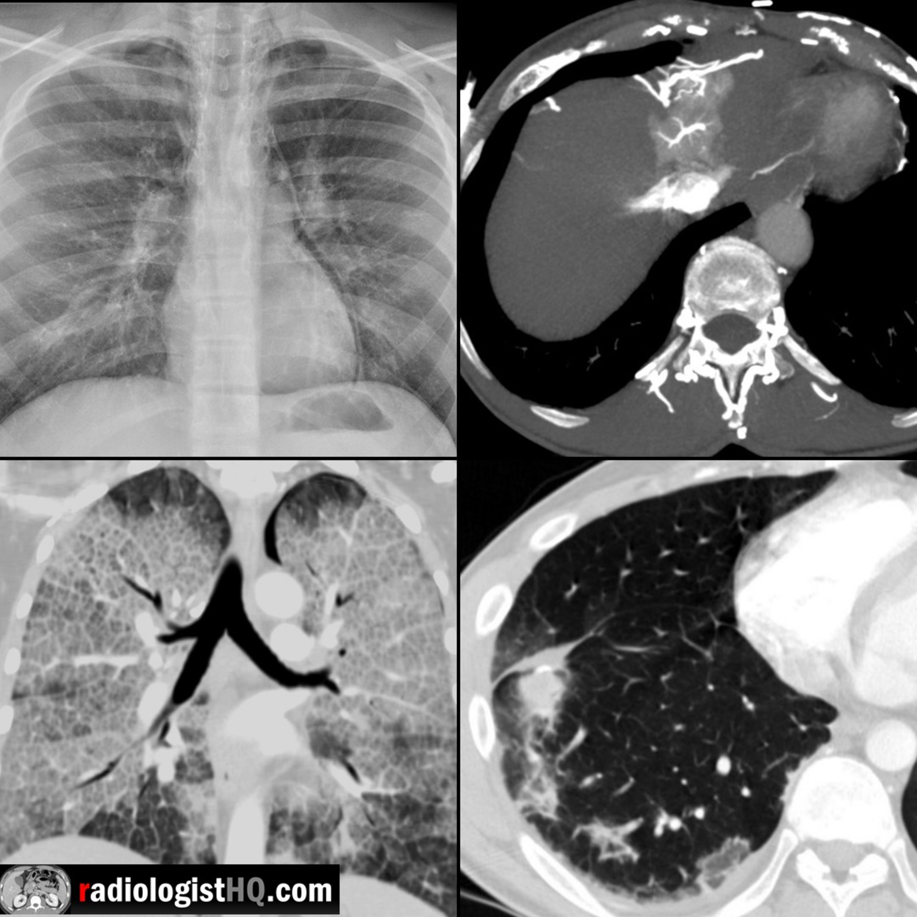 Radiology Lectures | Radquarters5 Cases in 5 Minutes: Thoracic #1Join us in this interactive video lecture as we present a total of 5 unknown thoracic imaging cases followed by a diagnosis reveal and key teaching points after each case, all in about 5(ish) minutes!
Click the YouTube Community tab or follow on social media for bonus teaching material posted throughout the week!
Website: https://radiologisthq.com/
Instagram: https://www.instagram.com/radiologistHQ/
Facebook: https://www.facebook.com/radiologistHeadQuarters/
Twitter: https://twitter.com/radiologistHQ
Reddit: https://www.reddit.com/user/radiologistHQ/
2019-03-2809 min
 Radiology Lectures | RadquartersHepatic Hemangioma: Pitfalls & Mimics, Part IIIn this video lecture, we focus on variants and malignant mimics of hemangioma and discuss how to characterize these masses on ultrasound, CT and MRI.
Key points include:
The ultrasound “target” sign is typical for hepatic metastases and appears as a lesion with a hypoechoic periphery and echogenic center.
Hemangiomas in a fatty liver may appear hypoechoic and mimic a more serious tumor but can be definitively characterized with MRI.
Sclerosed or hyalinized hemangiomas contain fibrous tissue and therefore have variable enhancement and diminished T2 hyperintensity.
Hemangiomas can be associated with arterioportal shunts and may be surr...2019-03-2119 min
 Radiology Lectures | RadquartersHepatic Hemangioma: Pitfalls & Mimics, Part IIn this video lecture, we discuss tips and tricks to diagnose everybody’s favorite hepatic tumor on CT, MRI and ultrasound.
Key points include:
Hemangioma is the most common benign hepatic tumor, and it is more common in females.
These tumors are usually asymptomatic and typically require no treatment, but can rarely cause pain, rupture if large, or cause Kasabach-Merritt syndrome.
On nonenhanced CT, hemangiomas will be hypodense to liver parenchyma and homogeneously isodense to the blood pool.
There are three major enhancement patterns for typical hemangiomas, and all patterns will show persistent delayed enhancement without co...2019-03-1415 min
 Radiology Lectures | Radquarters5 Cases in 5 Minutes: Ultrasound #5Join us in this interactive video lecture as we present a total of 5 unknown cases followed by a diagnosis reveal and key teaching points after each case, all in about 5(ish) minutes!
The post 5 Cases in 5 Minutes: Ultrasound #5 appeared first on Radquarters.
2019-03-0707 min
 Radiology Lectures | Radquarters5 Cases in 5 Minutes: Ultrasound #4Join us in this interactive lecture as we present a total of 5 unknown cases followed by a diagnosis reveal and key teaching points after each case, all in about 5(ish) minutes!
The post 5 Cases in 5 Minutes: Ultrasound #4 appeared first on Radquarters.
2019-02-2807 min
 Radiology Lectures | RadquartersIntroduction to Multiphase CT & MRI of the LiverIn this video lecture, we review the appearance of the liver on multiphase CT & MRI. A basic approach to image interpretation is presented with pitfalls to avoid.
Key points include:
The three major liver postcontrast phases include the late hepatic arterial phase, portal venous phase, and delayed/equilibrium phases.
The hepatic artery enhances first, followed by the portal veins, then the hepatic veins along with the hepatic parenchyma.
An ideal late hepatic arterial phase sequence will have both hepatic artery and portal vein enhancement with no hepatic vein enhancement.
The late hepatic arterial phase occurs at...2019-02-2116 min
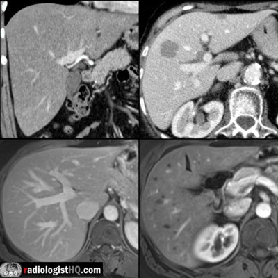 Radiology Lectures | RadquartersIntroduction to Multiphase CT & MRI of the LiverIn this video lecture, we review the appearance of the liver on multiphase CT & MRI. A basic approach to image interpretation is presented with pitfalls to avoid.
Key points include:
The three major liver postcontrast phases include the late hepatic arterial phase, portal venous phase, and delayed/equilibrium phases.
The hepatic artery enhances first, followed by the portal veins, then the hepatic veins along with the hepatic parenchyma.
An ideal late hepatic arterial phase sequence will have both hepatic artery and portal vein enhancement with no hepatic vein enhancement.
The late hepatic arterial phase occurs at a...2019-02-2116 min
 Radiology Lectures | Radquarters5 Cases in 5 Minutes: Ultrasound #3Join us in this interactive lecture as we present a total of 5 unknown cases followed by a diagnosis reveal and key teaching points after each case, all in about 5(ish) minutes!
The post 5 Cases in 5 Minutes: Ultrasound #3 appeared first on Radquarters.
2019-02-1407 min
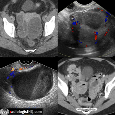 Radiology Lectures | RadquartersImaging of Pelvic Inflammatory Disease, Part IIn this video lecture, we discuss the ultrasound and computed tomography appearance of pelvic inflammatory disease (PID).
Topics include:
1) Early findings of PID, including haziness of pelvic fat, salpingitis, oophoritis, and endometritis.
2) Importance of identifying dilated fallopian tubes by the characteristic “C” or “S” shape that they exhibit, as well as the presence of the “cogwheel” sign.
3) Differentiating tubo-ovarian abscess (TOA) from tubo-ovarian complex (TOC), and the associated treatment implications.
4) Delineation of the typical posterior, dependent position of pyosalpinges and TOAs, as well as the associated anterior displacement of the broad li...2016-12-1409 min
 Writing ExcusesWriting Excuses 10.51: Q&A on Showing Your Work, with Daniel José OlderDaniel José Older joins us for a Q&A on showing your work around. Here are the questions, which were submitted by attendees at the Out of Excuses workshop: What's the best way to meet editors and agents at conventions? How do you write a good query letter? What do you mention as credentials in your query letter? You didn't cover self publishing at all this month. Self publishing is legit, right? Can you submit the same work to more than one agent or editor at a time? Can you re-submit a revised work to an agent who previously rej...2015-12-2122 min
 Season 10 – Writing ExcusesWriting Excuses 10.51: Q&A on Showing Your Work, with Daniel José OlderDaniel José Older joins us for a Q&A on showing your work around. Here are the questions, which were submitted by attendees at the Out of Excuses workshop: What’s the best way to meet editors and agents at conventions? How do you write a good query letter? What do you mention as credentials in your … Continue reading Writing Excuses 10.51: Q&A on Showing Your Work, with Daniel José Older →2015-12-2122 min
 2015 – Writing ExcusesWriting Excuses 10.51: Q&A on Showing Your Work, with Daniel José OlderDaniel José Older joins us for a Q&A on showing your work around. Here are the questions, which were submitted by attendees at the Out of Excuses workshop: What’s the best way to meet editors and agents at conventions? How do you write a good query letter? What do you mention as credentials in your … Continue reading Writing Excuses 10.51: Q&A on Showing Your Work, with Daniel José Older →2015-12-2122 min
 The Magic of Eyri PodcastAdapting a Novel to a Podcast (with Mary Robinette Kowal)This is audio from a panel discussion at Penguicon 2010 I was a part of, called “Adapting Your Novel to a Podcast.” Joining me on the panel is author, puppeteer and voice actor Mary Robinette Kowal. I recorded this live with my portable recorder placed between us, and I did what I could in GarageBand to boost the audio (and even out the levels), but a heads up that it is not as loud as I would of liked.
I really enjoyed this panel and Mary is just awesome. We approach podcasting from two different angles, with her bein...2010-05-0500 min