Shows

Medicine and ImagingCOVID-19: VERDADES E DÚVIDAS. ATUALIZAÇÃO EM SETEMBRO DE 2021. PARTE V: COVID-19 PÓS-AGUDAREFERÊNCIAS1.Pontone G, Scafuri S, Mancini ME, Agalbato C, Guglielmo M, Baggiano A, et al. Role of computed tomography in COVID-19. J Cardiovasc Comput Tomogr. 2021;15(1):27-36.2.Cereser L, Da Re J, Zuiani C, Girometti R. Chest high-resolution computed tomography is associated to short-time progression to severe disease in patients with COVID-19 pneumonia. Clin Imaging. 2021;70:61-6.3.Hochhegger B, Mandelli NS, Stuker G, Meirelles GSP, Zanon M, Mohammed TL, et al. Coronavirus Disease 2019 (COVID-19) Pneumonia Presentations in Chest Computed Tomography: A Pictorial Review. Curr Probl Diagn Radiol. 2021;50(3):436-42.4.Besutti G, Ottone M, Fasano T, Pattacini P, Iotti V...
2021-09-2702 min
Medicine and ImagingCOVID-19: VERDADES E DÚVIDAS. ATUALIZAÇÃO DE SETEMBRO DE 2021. PARTE IV: DOENÇA TROMBOEMBÓLICAREFERÊNCIAS1.Pontone G, Scafuri S, Mancini ME, Agalbato C, Guglielmo M, Baggiano A, et al. Role of computed tomography in COVID-19. J Cardiovasc Comput Tomogr. 2021;15(1):27-36.2.Cereser L, Da Re J, Zuiani C, Girometti R. Chest high-resolution computed tomography is associated to short-time progression to severe disease in patients with COVID-19 pneumonia. Clin Imaging. 2021;70:61-6.3.Hochhegger B, Mandelli NS, Stuker G, Meirelles GSP, Zanon M, Mohammed TL, et al. Coronavirus Disease 2019 (COVID-19) Pneumonia Presentations in Chest Computed Tomography: A Pictorial Review. Curr Probl Diagn Radiol. 2021;50(3):436-42.4.Besutti G, Ottone M, Fasano T, Pattacini P, Iotti V...
2021-09-2701 min
Medicine and ImagingCOVID-19: VERDADES E DÚVIDAS. ATUALIZAÇÃO DE SETEMBRO DE 2021. PARTE III: COMPARAÇÕES ENTRE A TC E HISTOPATOLOGIAREFERÊNCIAS1.Pontone G, Scafuri S, Mancini ME, Agalbato C, Guglielmo M, Baggiano A, et al. Role of computed tomography in COVID-19. J Cardiovasc Comput Tomogr. 2021;15(1):27-36.2.Cereser L, Da Re J, Zuiani C, Girometti R. Chest high-resolution computed tomography is associated to short-time progression to severe disease in patients with COVID-19 pneumonia. Clin Imaging. 2021;70:61-6.3.Hochhegger B, Mandelli NS, Stuker G, Meirelles GSP, Zanon M, Mohammed TL, et al. Coronavirus Disease 2019 (COVID-19) Pneumonia Presentations in Chest Computed Tomography: A Pictorial Review. Curr Probl Diagn Radiol. 2021;50(3):436-42.4.Besutti G, Ottone M, Fasano T, Pattacini P, Iotti V...
2021-09-2702 min
Medicine and ImagingCOVID-19: VERDADES E DÚVIDAS. ATUALIZAÇÃO DE SETEMBRO DE 2021. PARTE II: PROTOCOLOS DE LEITURA DA RSNA E O CORADSREFERÊNCIAS1.Pontone G, Scafuri S, Mancini ME, Agalbato C, Guglielmo M, Baggiano A, et al. Role of computed tomography in COVID-19. J Cardiovasc Comput Tomogr. 2021;15(1):27-36.2.Cereser L, Da Re J, Zuiani C, Girometti R. Chest high-resolution computed tomography is associated to short-time progression to severe disease in patients with COVID-19 pneumonia. Clin Imaging. 2021;70:61-6.3.Hochhegger B, Mandelli NS, Stuker G, Meirelles GSP, Zanon M, Mohammed TL, et al. Coronavirus Disease 2019 (COVID-19) Pneumonia Presentations in Chest Computed Tomography: A Pictorial Review. Curr Probl Diagn Radiol. 2021;50(3):436-42.4.Besutti G, Ottone M, Fasano T, Pattacini P, Iotti V...
2021-09-2702 min
Medicine and ImagingCOVID-19: VERDADES E DÚVIDAS. ATUALIZAÇÃO EM SETEMBRO DE 2021. - PARTE I: O PAPEL DA TC E RX NA COVID-19REFERÊNCIAS1.Pontone G, Scafuri S, Mancini ME, Agalbato C, Guglielmo M, Baggiano A, et al. Role of computed tomography in COVID-19. J Cardiovasc Comput Tomogr. 2021;15(1):27-36.2.Cereser L, Da Re J, Zuiani C, Girometti R. Chest high-resolution computed tomography is associated to short-time progression to severe disease in patients with COVID-19 pneumonia. Clin Imaging. 2021;70:61-6.3.Hochhegger B, Mandelli NS, Stuker G, Meirelles GSP, Zanon M, Mohammed TL, et al. Coronavirus Disease 2019 (COVID-19) Pneumonia Presentations in Chest Computed Tomography: A Pictorial Review. Curr Probl Diagn Radiol. 2021;50(3):436-42.4.Besutti G, Ottone M, Fasano T, Pattacini P, Iotti V...
2021-09-2705 min
Medicine and ImagingLESÕES DISTAIS DO BÍCEPS BRAQUIAL PARTE IIREFERENCES 1.Kulshreshtha R, Singh R, Sinha J, Hall S. Anatomy of the distal biceps brachii tendon and its clinical relevance. Clin Orthop Relat Res. 2007;456:117-20.2.Eames MH, Bain GI, Fogg QA, van Riet RP. Distal biceps tendon anatomy: a cadaveric study. J Bone Joint Surg Am. 2007;89(5):1044-9.3.Athwal GS, Steinmann SP, Rispoli DM. The distal biceps tendon: footprint and relevant clinical anatomy. J Hand Surg Am. 2007;32(8):1225-9.4.Konschake M, Stofferin H, Moriggl B. Ultrasound visualization of an underestimated structure: the bicipital aponeurosis. Surg Radiol Anat. 2017;39(12):1317-22.5.Snoeck O, Lefevre P, Sprio E, Beslay R...
2021-04-2504 min
Medicine and ImagingLESÕES DISTAIS DO BÍCEPS BRAQUIAL PARTE IREFERENCES 1.Kulshreshtha R, Singh R, Sinha J, Hall S. Anatomy of the distal biceps brachii tendon and its clinical relevance. Clin Orthop Relat Res. 2007;456:117-20.2.Eames MH, Bain GI, Fogg QA, van Riet RP. Distal biceps tendon anatomy: a cadaveric study. J Bone Joint Surg Am. 2007;89(5):1044-9.3.Athwal GS, Steinmann SP, Rispoli DM. The distal biceps tendon: footprint and relevant clinical anatomy. J Hand Surg Am. 2007;32(8):1225-9.4.Konschake M, Stofferin H, Moriggl B. Ultrasound visualization of an underestimated structure: the bicipital aponeurosis. Surg Radiol Anat. 2017;39(12):1317-22.5.Snoeck O, Lefevre P, Sprio E, Beslay R...
2021-04-2504 min
Medicine and ImagingBICEPS BRACHIALIS DISTAL TEARSREFERENCES 1.Kulshreshtha R, Singh R, Sinha J, Hall S. Anatomy of the distal biceps brachii tendon and its clinical relevance. Clin Orthop Relat Res. 2007;456:117-20.2.Eames MH, Bain GI, Fogg QA, van Riet RP. Distal biceps tendon anatomy: a cadaveric study. J Bone Joint Surg Am. 2007;89(5):1044-9.3.Athwal GS, Steinmann SP, Rispoli DM. The distal biceps tendon: footprint and relevant clinical anatomy. J Hand Surg Am. 2007;32(8):1225-9.4.Konschake M, Stofferin H, Moriggl B. Ultrasound visualization of an underestimated structure: the bicipital aponeurosis. Surg Radiol Anat. 2017;39(12):1317-22.5.Snoeck O, Lefevre P, Sprio E, Beslay R...
2021-04-2507 min
Medicine and ImagingDoença Torácicas Relacionada ao Amianto Parte IIReferências1.Mogami R, Marchiori E, Albuquerque HA, Ribeiro P, Capone D. Correlação entre Radiografia Convencional e Tomografia Computadorizada de Alta Resolução de Tórax em Trabalhadores da Indústria Têxtil do Asbesto. Rev Imagem 2001;23(4):233-8.2.Kim KI, Kim CW, Lee MK, Lee KS, Park CK, Choi SJ, et al. Imaging of occupational lung disease. Radiographics. 2001;21(6):1371-91.3.Oury T; Sporn TA; Roggli VL. Pathology of Asbestos-Associated Diseases. Third ed: Springer; 2014.4.Copley SJ, Wells AU, Sivakumaran P, Rubens MB, Lee YC, Desai SR, et al. Asbestosis and idiopathic pulmonary fibrosis: comparison of thin-section CT features...
2021-04-1903 min
Medicine and ImagingDoenças Torácicas Relacionadas ao Amianto Parte IReferências1.Mogami R, Marchiori E, Albuquerque HA, Ribeiro P, Capone D. Correlação entre Radiografia Convencional e Tomografia Computadorizada de Alta Resolução de Tórax em Trabalhadores da Indústria Têxtil do Asbesto. Rev Imagem 2001;23(4):233-8.2.Kim KI, Kim CW, Lee MK, Lee KS, Park CK, Choi SJ, et al. Imaging of occupational lung disease. Radiographics. 2001;21(6):1371-91.3.Oury T; Sporn TA; Roggli VL. Pathology of Asbestos-Associated Diseases. Third ed: Springer; 2014.4.Copley SJ, Wells AU, Sivakumaran P, Rubens MB, Lee YC, Desai SR, et al. Asbestosis and idiopathic pulmonary fibrosis: comparison of thin-section CT features...
2021-04-1907 min
Medicine and ImagingTHE ROTATOR CABLEReferences1.Nozaki T, Nimura A, Fujishiro H, Mochizuki T, Yamaguchi K, Kato R, et al. The anatomic relationship between the morphology of the greater tubercle of the humerus and the insertion of the infraspinatus tendon. J Shoulder Elbow Surg. 2015;24(4):555-60.2.Mochizuki T, Sugaya H, Uomizu M, Maeda K, Matsuki K, Sekiya I, et al. Humeral insertion of the supraspinatus and infraspinatus. New anatomical findings regarding the footprint of the rotator cuff. Surgical technique. J Bone Joint Surg Am. 2009;91 Suppl 2 Pt 1:1-7.3.Chang EY, Chung CB. Current concepts on imaging diagnosis of rotator cuff disease. Semin Musculoskelet...
2021-02-0508 min
Medicine and ImagingCRURAL FASCIAReferences1.Stecco C, Cappellari A, Macchi V, Porzionato A, Morra A, Berizzi A, et al. The paratendineous tissues: an anatomical study of their role in the pathogenesis of tendinopathy. Surg Radiol Anat. 2014;36(6):561-72.2.Silberman MR AK. Crural Fascia Injuries and Thickening on Musculoskeletal Ultrasound in Athletes with Calf Pain, Calf “Strains”, Exertional Compartiment Syndrome and Butulinum Toxin Injections. The Journal of Sports Medicine and Performance. 2017.3.Stecco C. Functional Atlas of the Human Fascial System: Churchill Livingstone Elsevier; 2015.4.Morton S, Chan O, Webborn N, Pritchard M, Morrissey D. Tears of the fascia cruris demonstrate characteristic sonographic features: a ca...
2021-01-2605 min
Medicine and ImagingACROMIOCLAVICULAR JOINT - PART IIReferences1.Flores DV, Goes PK, Gomez CM, Umpire DF, Pathria MN. Imaging of the Acromioclavicular Joint: Anatomy, Function, Pathologic Features, and Treatment. Radiographics. 2020;40(5):1355-82.2.Ha AS, Petscavage-Thomas JM, Tagoylo GH. Acromioclavicular joint: the other joint in the shoulder. AJR Am J Roentgenol. 2014;202(2):375-85.3.Goes PCK, Pathria MN. Radiographic/MR Imaging Correlation of the Shoulder. Magn Reson Imaging Clin N Am. 2019;27(4):575-85.4.Faruch Bilfeld M, Lapegue F, Chiavassa Gandois H, Bayol MA, Bonnevialle N, Sans N. Ultrasound of the coracoclavicular ligaments in the acute phase of an acromioclavicular disjonction: Comparison of radiographic, ultrasound and MRI findings. Eur...
2021-01-2106 min
Medicine and ImagingACROMIOCLAVICULAR JOINT - PART IReferences1.Flores DV, Goes PK, Gomez CM, Umpire DF, Pathria MN. Imaging of the Acromioclavicular Joint: Anatomy, Function, Pathologic Features, and Treatment. Radiographics. 2020;40(5):1355-82.2.Ha AS, Petscavage-Thomas JM, Tagoylo GH. Acromioclavicular joint: the other joint in the shoulder. AJR Am J Roentgenol. 2014;202(2):375-85.3.Goes PCK, Pathria MN. Radiographic/MR Imaging Correlation of the Shoulder. Magn Reson Imaging Clin N Am. 2019;27(4):575-85.4.Faruch Bilfeld M, Lapegue F, Chiavassa Gandois H, Bayol MA, Bonnevialle N, Sans N. Ultrasound of the coracoclavicular ligaments in the acute phase of an acromioclavicular disjonction: Comparison of radiographic, ultrasound and MRI findings. Eur...
2021-01-2105 min
Medicine and ImagingMUSCULAR ANATOMY OF THE SOLE OF THE FOOTReferences:1.Sarrafian SK. Sarrafian’s Anatomy of theFoot and Ankle Descriptive, Topographic, Functional. third ed2011.2.Edama M, Takabayashi T, Inai T, Kikumoto T, Hirabayashi R, Ito W, et al. The relationships between the quadratus plantae and the flexor digitorum longus and the flexor hallucis longus. Surg Radiol Anat. 2019;41(6):689-92.3.Willegger M, Seyidova N, Schuh R, Windhager R, Hirtler L. The tibialis posterior tendon footprint: an anatomical dissection study. J Foot Ankle Res. 2020;13(1):25.4.Hallinan J, Wang W, Pathria MN, Smitaman E, Huang BK. The peroneus longus muscle and tendon: a review of its anatomy and pa...
2021-01-1108 min
Medicine and ImagingHallux sesamoid complex imaging a practical diagnostic approach SKELETAL RADIOLOGY 2020A summary of the review paper of Skeletal Radiology (2020) 49:1889–1901https://doi.org/10.1007/s00256-020-03507-8
2021-01-0605 min
Medicine and ImagingMUSCLE INJURIES PART XVI - ADDUCTOR INJURIESReferences1. Flores DV, Mejia Gomez C, Estrada-Castrillon M, Smitaman E, Pathria MN. MR Imaging of Muscle Trauma: Anatomy, Biomechanics, Pathophysiology, and Imaging Appearance. Radiographics. 2018;38(1):124-48.2. Pathria M. MRI traumatic changes 2009 (Radiology Assistant)3. Study Group of the M, Tendon System from the Spanish Society of Sports T, Balius R, Blasi M, Pedret C, Alomar X, et al. A Histoarchitectural Approach to Skeletal Muscle Injury: Searching for a Common Nomenclature. Orthop J Sports Med. 2020;8(3):2325967120909090.4. Balius R, Alomar X, Pedret C, Blasi M, Rodas G, Pruna R, et al. Role of the Extracellular Matrix in Muscle Injuries: Histoarchitectural Considerations...
2021-01-0304 min
Medicine and ImagingMUSCLE INJURIES PART XV - RECTUS FEMORIS' TEARSReferences1. Flores DV, Mejia Gomez C, Estrada-Castrillon M, Smitaman E, Pathria MN. MR Imaging of Muscle Trauma: Anatomy, Biomechanics, Pathophysiology, and Imaging Appearance. Radiographics. 2018;38(1):124-48.2. Pathria M. MRI traumatic changes 2009 (Radiology Assistant)3. Study Group of the M, Tendon System from the Spanish Society of Sports T, Balius R, Blasi M, Pedret C, Alomar X, et al. A Histoarchitectural Approach to Skeletal Muscle Injury: Searching for a Common Nomenclature. Orthop J Sports Med. 2020;8(3):2325967120909090.4. Balius R, Alomar X, Pedret C, Blasi M, Rodas G, Pruna R, et al. Role of the Extracellular Matrix in Muscle Injuries: Histoarchitectural Considerations...
2021-01-0304 min
Medicine and ImagingMUSCLE INJURIES PART XIV (HAMSTRING INJURIES PART III)References1. Flores DV, Mejia Gomez C, Estrada-Castrillon M, Smitaman E, Pathria MN. MR Imaging of Muscle Trauma: Anatomy, Biomechanics, Pathophysiology, and Imaging Appearance. Radiographics. 2018;38(1):124-48.2. Pathria M. MRI traumatic changes 2009 (Radiology Assistant)3. Study Group of the M, Tendon System from the Spanish Society of Sports T, Balius R, Blasi M, Pedret C, Alomar X, et al. A Histoarchitectural Approach to Skeletal Muscle Injury: Searching for a Common Nomenclature. Orthop J Sports Med. 2020;8(3):2325967120909090.4. Balius R, Alomar X, Pedret C, Blasi M, Rodas G, Pruna R, et al. Role of the Extracellular Matrix in Muscle Injuries: Histoarchitectural Considerations...
2021-01-0301 min
Medicine and ImagingMUSCLE INJURIES PART XIII (HAMSTRING INJURIES PART II)References1. Flores DV, Mejia Gomez C, Estrada-Castrillon M, Smitaman E, Pathria MN. MR Imaging of Muscle Trauma: Anatomy, Biomechanics, Pathophysiology, and Imaging Appearance. Radiographics. 2018;38(1):124-48.2. Pathria M. MRI traumatic changes 2009 (Radiology Assistant)3. Study Group of the M, Tendon System from the Spanish Society of Sports T, Balius R, Blasi M, Pedret C, Alomar X, et al. A Histoarchitectural Approach to Skeletal Muscle Injury: Searching for a Common Nomenclature. Orthop J Sports Med. 2020;8(3):2325967120909090.4. Balius R, Alomar X, Pedret C, Blasi M, Rodas G, Pruna R, et al. Role of the Extracellular Matrix in Muscle Injuries: Histoarchitectural Considerations...
2021-01-0305 min
Medicine and ImagingMUSCLE INJURIES PART XII (HAMSTRING INJURIES PART I)References1. Flores DV, Mejia Gomez C, Estrada-Castrillon M, Smitaman E, Pathria MN. MR Imaging of Muscle Trauma: Anatomy, Biomechanics, Pathophysiology, and Imaging Appearance. Radiographics. 2018;38(1):124-48.2. Pathria M. MRI traumatic changes 2009 (Radiology Assistant)3. Study Group of the M, Tendon System from the Spanish Society of Sports T, Balius R, Blasi M, Pedret C, Alomar X, et al. A Histoarchitectural Approach to Skeletal Muscle Injury: Searching for a Common Nomenclature. Orthop J Sports Med. 2020;8(3):2325967120909090.4. Balius R, Alomar X, Pedret C, Blasi M, Rodas G, Pruna R, et al. Role of the Extracellular Matrix in Muscle Injuries: Histoarchitectural Considerations...
2021-01-0305 min
Medicine and ImagingMUSCLE INJURIES PART XI (CALF INJURIES PART II)References1. Flores DV, Mejia Gomez C, Estrada-Castrillon M, Smitaman E, Pathria MN. MR Imaging of Muscle Trauma: Anatomy, Biomechanics, Pathophysiology, and Imaging Appearance. Radiographics. 2018;38(1):124-48.2. Pathria M. MRI traumatic changes 2009 (Radiology Assistant)3. Study Group of the M, Tendon System from the Spanish Society of Sports T, Balius R, Blasi M, Pedret C, Alomar X, et al. A Histoarchitectural Approach to Skeletal Muscle Injury: Searching for a Common Nomenclature. Orthop J Sports Med. 2020;8(3):2325967120909090.4. Balius R, Alomar X, Pedret C, Blasi M, Rodas G, Pruna R, et al. Role of the Extracellular Matrix in Muscle Injuries: Histoarchitectural Considerations...
2021-01-0304 min
Medicine and ImagingMUSCLE INJURIES PART X (CALF INJURIES PART I)References1. Flores DV, Mejia Gomez C, Estrada-Castrillon M, Smitaman E, Pathria MN. MR Imaging of Muscle Trauma: Anatomy, Biomechanics, Pathophysiology, and Imaging Appearance. Radiographics. 2018;38(1):124-48.2. Pathria M. MRI traumatic changes 2009 (Radiology Assistant)3. Study Group of the M, Tendon System from the Spanish Society of Sports T, Balius R, Blasi M, Pedret C, Alomar X, et al. A Histoarchitectural Approach to Skeletal Muscle Injury: Searching for a Common Nomenclature. Orthop J Sports Med. 2020;8(3):2325967120909090.4. Balius R, Alomar X, Pedret C, Blasi M, Rodas G, Pruna R, et al. Role of the Extracellular Matrix in Muscle Injuries: Histoarchitectural Considerations...
2021-01-0304 min
Medicine and ImagingMUSCLE INJURIES PART IX - REPAIRReferences1. Flores DV, Mejia Gomez C, Estrada-Castrillon M, Smitaman E, Pathria MN. MR Imaging of Muscle Trauma: Anatomy, Biomechanics, Pathophysiology, and Imaging Appearance. Radiographics. 2018;38(1):124-48.2. Pathria M. MRI traumatic changes 2009 (Radiology Assistant)3. Study Group of the M, Tendon System from the Spanish Society of Sports T, Balius R, Blasi M, Pedret C, Alomar X, et al. A Histoarchitectural Approach to Skeletal Muscle Injury: Searching for a Common Nomenclature. Orthop J Sports Med. 2020;8(3):2325967120909090.4. Balius R, Alomar X, Pedret C, Blasi M, Rodas G, Pruna R, et al. Role of the Extracellular Matrix in Muscle Injuries: Histoarchitectural Considerations...
2021-01-0301 min
Medicine and ImagingMUSCLE INJURIES PART VIII - FOLLOW-UP WITH USReferences1. Flores DV, Mejia Gomez C, Estrada-Castrillon M, Smitaman E, Pathria MN. MR Imaging of Muscle Trauma: Anatomy, Biomechanics, Pathophysiology, and Imaging Appearance. Radiographics. 2018;38(1):124-48.2. Pathria M. MRI traumatic changes 2009 (Radiology Assistant)3. Study Group of the M, Tendon System from the Spanish Society of Sports T, Balius R, Blasi M, Pedret C, Alomar X, et al. A Histoarchitectural Approach to Skeletal Muscle Injury: Searching for a Common Nomenclature. Orthop J Sports Med. 2020;8(3):2325967120909090.4. Balius R, Alomar X, Pedret C, Blasi M, Rodas G, Pruna R, et al. Role of the Extracellular Matrix in Muscle Injuries: Histoarchitectural Considerations...
2021-01-0302 min
Medicine and ImagingMUSCLE INJURIES PART VII - THE BRITISH CLASSIFICATION AND THE BARCELONA-ASPETAR-DUKE CLASSIFICATIONReferences1. Flores DV, Mejia Gomez C, Estrada-Castrillon M, Smitaman E, Pathria MN. MR Imaging of Muscle Trauma: Anatomy, Biomechanics, Pathophysiology, and Imaging Appearance. Radiographics. 2018;38(1):124-48.2. Pathria M. MRI traumatic changes 2009 (Radiology Assistant)3. Study Group of the M, Tendon System from the Spanish Society of Sports T, Balius R, Blasi M, Pedret C, Alomar X, et al. A Histoarchitectural Approach to Skeletal Muscle Injury: Searching for a Common Nomenclature. Orthop J Sports Med. 2020;8(3):2325967120909090.4. Balius R, Alomar X, Pedret C, Blasi M, Rodas G, Pruna R, et al. Role of the Extracellular Matrix in Muscle Injuries: Histoarchitectural Considerations...
2021-01-0304 min
Medicine and ImagingMUSCLE INJURIES PART VI - THE BRITISH CLASSIFICATIONReferences1. Flores DV, Mejia Gomez C, Estrada-Castrillon M, Smitaman E, Pathria MN. MR Imaging of Muscle Trauma: Anatomy, Biomechanics, Pathophysiology, and Imaging Appearance. Radiographics. 2018;38(1):124-48.2. Pathria M. MRI traumatic changes 2009 (Radiology Assistant)3. Study Group of the M, Tendon System from the Spanish Society of Sports T, Balius R, Blasi M, Pedret C, Alomar X, et al. A Histoarchitectural Approach to Skeletal Muscle Injury: Searching for a Common Nomenclature. Orthop J Sports Med. 2020;8(3):2325967120909090.4. Balius R, Alomar X, Pedret C, Blasi M, Rodas G, Pruna R, et al. Role of the Extracellular Matrix in Muscle Injuries: Histoarchitectural Considerations...
2021-01-0204 min
Medicine and ImagingMUSCLE INJURIES PART V - THE MUNICH CLASSIFICATIONReferences1. Flores DV, Mejia Gomez C, Estrada-Castrillon M, Smitaman E, Pathria MN. MR Imaging of Muscle Trauma: Anatomy, Biomechanics, Pathophysiology, and Imaging Appearance. Radiographics. 2018;38(1):124-48.2. Pathria M. MRI traumatic changes 2009 (Radiology Assistant)3. Study Group of the M, Tendon System from the Spanish Society of Sports T, Balius R, Blasi M, Pedret C, Alomar X, et al. A Histoarchitectural Approach to Skeletal Muscle Injury: Searching for a Common Nomenclature. Orthop J Sports Med. 2020;8(3):2325967120909090.4. Balius R, Alomar X, Pedret C, Blasi M, Rodas G, Pruna R, et al. Role of the Extracellular Matrix in Muscle Injuries: Histoarchitectural Considerations...
2021-01-0204 min
Medicine and ImagingMUSCLE INJURIES PART IV - RETURN TO PLAY AND STAGINGReferences1. Flores DV, Mejia Gomez C, Estrada-Castrillon M, Smitaman E, Pathria MN. MR Imaging of Muscle Trauma: Anatomy, Biomechanics, Pathophysiology, and Imaging Appearance. Radiographics. 2018;38(1):124-48.2. Pathria M. MRI traumatic changes 2009 (Radiology Assistant)3. Study Group of the M, Tendon System from the Spanish Society of Sports T, Balius R, Blasi M, Pedret C, Alomar X, et al. A Histoarchitectural Approach to Skeletal Muscle Injury: Searching for a Common Nomenclature. Orthop J Sports Med. 2020;8(3):2325967120909090.4. Balius R, Alomar X, Pedret C, Blasi M, Rodas G, Pruna R, et al. Role of the Extracellular Matrix in Muscle Injuries: Histoarchitectural Considerations...
2021-01-0204 min
Medicine and ImagingMUSCLE INJURIES PART III - CONTUSION, LACERATION AND COMPARTMENTAL SYNDROMESReferences1. Flores DV, Mejia Gomez C, Estrada-Castrillon M, Smitaman E, Pathria MN. MR Imaging of Muscle Trauma: Anatomy, Biomechanics, Pathophysiology, and Imaging Appearance. Radiographics. 2018;38(1):124-48.2. Pathria M. MRI traumatic changes 2009 (Radiology Assistant)3. Study Group of the M, Tendon System from the Spanish Society of Sports T, Balius R, Blasi M, Pedret C, Alomar X, et al. A Histoarchitectural Approach to Skeletal Muscle Injury: Searching for a Common Nomenclature. Orthop J Sports Med. 2020;8(3):2325967120909090.4. Balius R, Alomar X, Pedret C, Blasi M, Rodas G, Pruna R, et al. Role of the Extracellular Matrix in Muscle Injuries: Histoarchitectural Considerations...
2021-01-0203 min
Medicine and ImagingMUSCLE INJURIES PART II - TEARS AND DOMSReferences1.Flores DV, Mejia Gomez C, Estrada-Castrillon M, Smitaman E, Pathria MN. MR Imaging of Muscle Trauma: Anatomy, Biomechanics, Pathophysiology, and Imaging Appearance. Radiographics. 2018;38(1):124-48.2.Pathria M. MRI traumatic changes 2009 [Available from: https://radiologyassistant.nl/musculoskeletal/muscle/mri-traumatic-changes.3.Study Group of the M, Tendon System from the Spanish Society of Sports T, Balius R, Blasi M, Pedret C, Alomar X, et al. A Histoarchitectural Approach to Skeletal Muscle Injury: Searching for a Common Nomenclature. Orthop J Sports Med. 2020;8(3):2325967120909090.4.Balius R, Alomar X, Pedret C, Blasi M, Rodas G, Pruna R, et al. Role of the Extracellular...
2021-01-0203 min
Medicine and ImagingMUSCLE INJURIES PART I - INTRODUCTION AND ANATOMYReferences1.Flores DV, Mejia Gomez C, Estrada-Castrillon M, Smitaman E, Pathria MN. MR Imaging of Muscle Trauma: Anatomy, Biomechanics, Pathophysiology, and Imaging Appearance. Radiographics. 2018;38(1):124-48.2.Pathria M. MRI traumatic changes 2009 [Available from: https://radiologyassistant.nl/musculoskeletal/muscle/mri-traumatic-changes.3.Study Group of the M, Tendon System from the Spanish Society of Sports T, Balius R, Blasi M, Pedret C, Alomar X, et al. A Histoarchitectural Approach to Skeletal Muscle Injury: Searching for a Common Nomenclature. Orthop J Sports Med. 2020;8(3):2325967120909090.4.Balius R, Alomar X, Pedret C, Blasi M, Rodas G, Pruna R, et al. Role of the Extracellular...
2021-01-0206 min
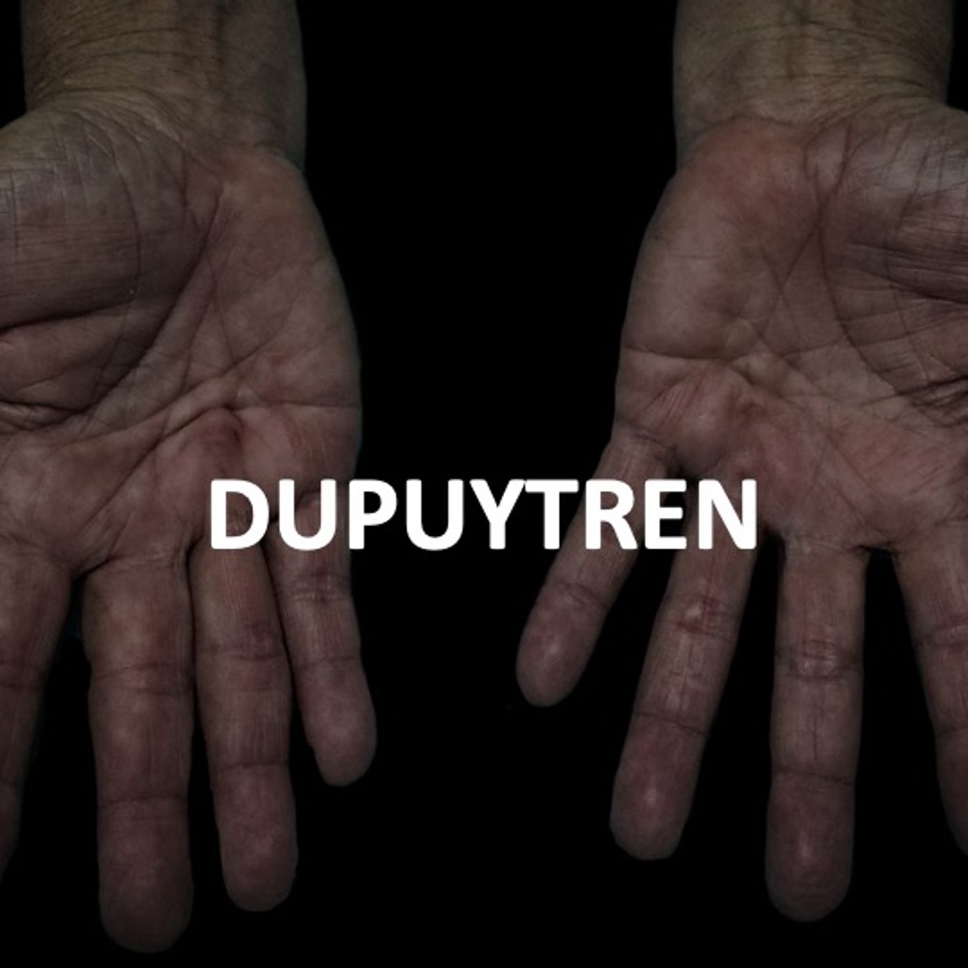
Medicine and ImagingDUPUYTREN1- Introdução2- Anatomia da aponeurose palmar3- Fisiopatologia4- Aspectos de imagemReferências:1.Créteur V, Madani A, Gosset N . Ultrasound imaging of Dupuytren’s contracture J Radiol. 2010;91:687-912.Morris G, Jacobson JA, Kalume Brigido M, et al. Ultrasound Features of Palmar Fibromatosis or Dupuytren Contracture. J Ultrasound Med. 2019;38(2):387-92.3.Grazina R, Teixeira S, Ramos R et al. Dupuytren’s disease: where do we stand? EFORT Open Rev. 2019;4:63-9.4.Balaban M, Cilengir AH, Idilman IS. Evaluation of Tendon Disorders With Ultrasonography and Elastography. J Ultrasound Med. 2020.5.Rubin D. Dupuy...
2020-12-0507 min
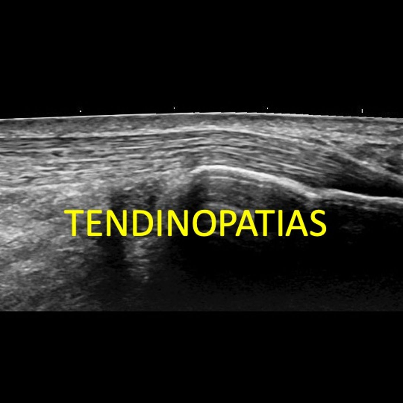
Medicine and ImagingTENDINOPATIAS PARTE III - TEORIA DO ICEBERG E EXAMES DE IMAGEMReferências1.Khan KM, Cook JL, Bonar F, Harcourt P, Astrom M. Histopathology of common tendinopathies. Update and implications for clinical management. Sports Med. 1999;27(6):393-408.2.Andarawis-Puri N, Flatow EL, Soslowsky LJ. Tendon basic science: Development, repair, regeneration, and healing. J Orthop Res. 2015;33(6):780-4.3.Ryan M, Bisset L, Newsham-West R. Should We Care About Tendon Structure? The Disconnect Between Structure and Symptoms in Tendinopathy. J Orthop Sports Phys Ther. 2015;45(11):823-5.4.Scott A, Backman LJ, Speed C. Tendinopathy: Update on Pathophysiology. J Orthop Sports Phys Ther. 2015;45(11):833-41.5.Abate M, Silbernagel KG, Siljeholm C, Di Iorio A, De A...
2020-11-2903 min
Medicine and ImagingTENDINOPATIAS PARTE II - TEORIA DO CONTINUUMReferências1.Khan KM, Cook JL, Bonar F, Harcourt P, Astrom M. Histopathology of common tendinopathies. Update and implications for clinical management. Sports Med. 1999;27(6):393-408.2.Andarawis-Puri N, Flatow EL, Soslowsky LJ. Tendon basic science: Development, repair, regeneration, and healing. J Orthop Res. 2015;33(6):780-4.3.Ryan M, Bisset L, Newsham-West R. Should We Care About Tendon Structure? The Disconnect Between Structure and Symptoms in Tendinopathy. J Orthop Sports Phys Ther. 2015;45(11):823-5.4.Scott A, Backman LJ, Speed C. Tendinopathy: Update on Pathophysiology. J Orthop Sports Phys Ther. 2015;45(11):833-41.5.Abate M, Silbernagel KG, Siljeholm C, Di Iorio A, De A...
2020-11-2904 min
Medicine and ImagingTENDINOPATIAS PARTE I - INTRODUÇÃOReferências1.Khan KM, Cook JL, Bonar F, Harcourt P, Astrom M. Histopathology of common tendinopathies. Update and implications for clinical management. Sports Med. 1999;27(6):393-408.2.Andarawis-Puri N, Flatow EL, Soslowsky LJ. Tendon basic science: Development, repair, regeneration, and healing. J Orthop Res. 2015;33(6):780-4.3.Ryan M, Bisset L, Newsham-West R. Should We Care About Tendon Structure? The Disconnect Between Structure and Symptoms in Tendinopathy. J Orthop Sports Phys Ther. 2015;45(11):823-5.4.Scott A, Backman LJ, Speed C. Tendinopathy: Update on Pathophysiology. J Orthop Sports Phys Ther. 2015;45(11):833-41.5.Abate M, Silbernagel KG, Siljeholm C, Di Iorio A, De A...
2020-11-2906 min
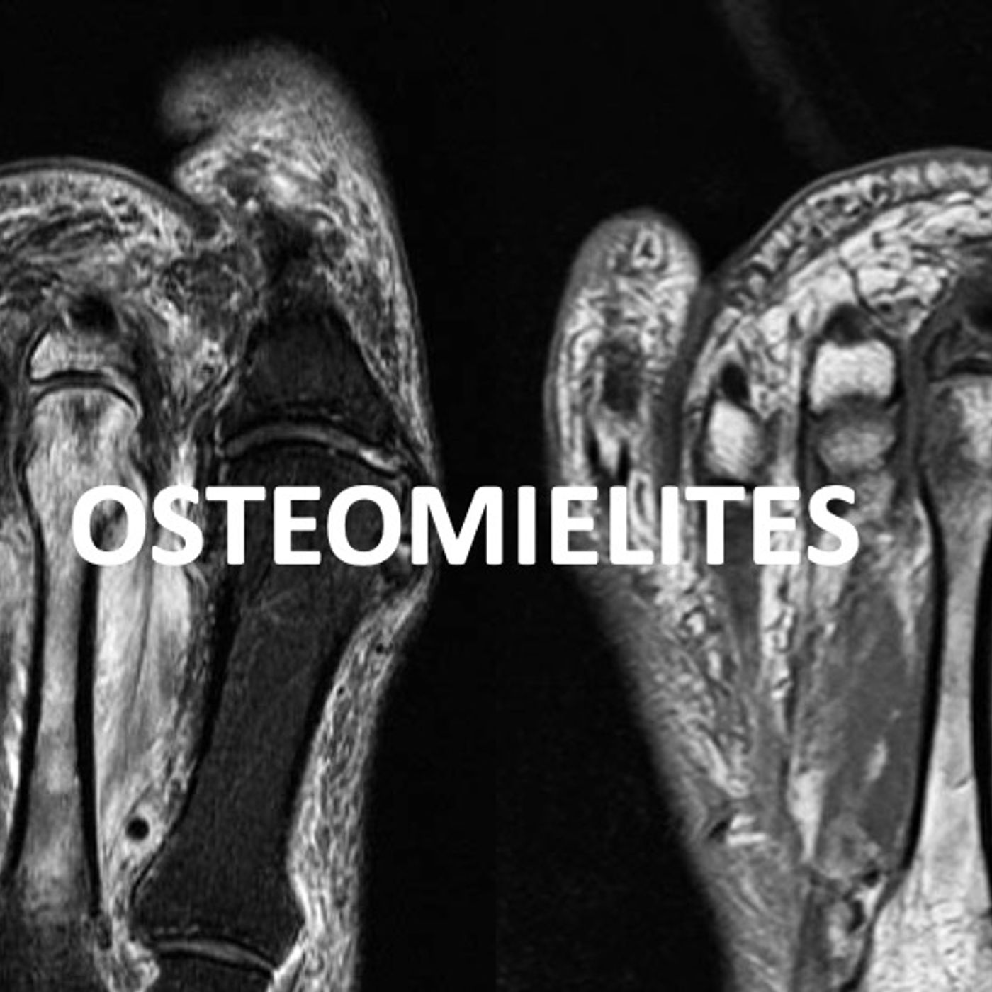
Medicine and ImagingOSTEOMIELITESAtualização de um podcast sobre "Osteomielites Agudas" publicaod em 6/7/2020
2020-11-2610 min
Medicine and ImagingFRATURASReferências1.Resnick D, Kransdorf MJ. Physical Injury: Concepts and Terminology. In: Bone and Joint Imaging. Terceira edição. Philadelphia. Elsevier Saunders. 2005.2.Greenspan A, Beltran J. Radiologic Evaluation of Trauma. In: Orthopedic Imaging A Practical Approach. Sexta edição. New York. Lippincott Williams & Wilkins. 2014.3.Narayanasamy S, Krishna S, Sathiadoss P, Althobaity W, Koujok K, Sheikh AM. Radiographic Review of Avulsion Fractures RadioGraphics Fundamentals | Online Presentation. Radiographics. 2018;38(5):1496-7.4.Thompson JC. Ciência Básica. In: Netter Atlas de Anatomia Ortopédica. Segunda edição. Rio de Janeiro. Elsevier Editora Ltda. 2012.5.Marshall RA, Mandell JC, Weaver MJ, Ferrone M...
2020-11-2313 min
Medicine and ImagingArtrite Reumatoide - Manifestações extra-articulares, padrões de acometimento, clasificação e emprego dos métodos de imagemReferências1.Filippucci E, Cipolletta E, Mashadi Mirza R, Carotti M, Giovagnoni A, Salaffi F, et al. Ultrasound imaging in rheumatoid arthritis. Radiol Med. 2019;124(11):1087-100.2.Rowbotham EL, Grainger AJ. Rheumatoid arthritis: ultrasound versus MRI. AJR Am J Roentgenol. 2011;197(3):541-6.3.Scott DL, Wolfe F, Huizinga TW. Rheumatoid arthritis. Lancet. 2010;376(9746):1094-108.4.Ngian GS. Rheumatoid arthritis. Aust Fam Physician. 2010;39(9):626-8.5.Sommer OJ, Kladosek A, Weiler V, Czembirek H, Boeck M, Stiskal M. Rheumatoid arthritis: a practical guide to state-of-the-art imaging, image interpretation, and clinical implications. Radiographics. 2005;25(2):381-98.6.Chang EY, Chen KC, Huang BK, Kavanaugh A. Adult Inflammatory A...
2020-11-1705 min
Medicine and ImagingArtrite Reumatoide - Manifestações ArticularesReferências1.Filippucci E, Cipolletta E, Mashadi Mirza R, Carotti M, Giovagnoni A, Salaffi F, et al. Ultrasound imaging in rheumatoid arthritis. Radiol Med. 2019;124(11):1087-100.2.Rowbotham EL, Grainger AJ. Rheumatoid arthritis: ultrasound versus MRI. AJR Am J Roentgenol. 2011;197(3):541-6.3.Scott DL, Wolfe F, Huizinga TW. Rheumatoid arthritis. Lancet. 2010;376(9746):1094-108.4.Ngian GS. Rheumatoid arthritis. Aust Fam Physician. 2010;39(9):626-8.5.Sommer OJ, Kladosek A, Weiler V, Czembirek H, Boeck M, Stiskal M. Rheumatoid arthritis: a practical guide to state-of-the-art imaging, image interpretation, and clinical implications. Radiographics. 2005;25(2):381-98.6.Chang EY, Chen KC, Huang BK, Kavanaugh A. Adult Inflammatory A...
2020-11-1708 min
Medicine and ImagingArtrite Reumatoide - IntroduçãoReferências1.Filippucci E, Cipolletta E, Mashadi Mirza R, Carotti M, Giovagnoni A, Salaffi F, et al. Ultrasound imaging in rheumatoid arthritis. Radiol Med. 2019;124(11):1087-100.2.Rowbotham EL, Grainger AJ. Rheumatoid arthritis: ultrasound versus MRI. AJR Am J Roentgenol. 2011;197(3):541-6.3.Scott DL, Wolfe F, Huizinga TW. Rheumatoid arthritis. Lancet. 2010;376(9746):1094-108.4.Ngian GS. Rheumatoid arthritis. Aust Fam Physician. 2010;39(9):626-8.5.Sommer OJ, Kladosek A, Weiler V, Czembirek H, Boeck M, Stiskal M. Rheumatoid arthritis: a practical guide to state-of-the-art imaging, image interpretation, and clinical implications. Radiographics. 2005;25(2):381-98.6.Chang EY, Chen KC, Huang BK, Kavanaugh A. Adult Inflammatory A...
2020-11-1704 min
Medicine and ImagingELBOW LIGAMENTS - PART II1- Introduction;2- Lateral Collateral Ligament Complex;3- Ulnar Collateral Ligament Complex;4- Valgus Extension Overload Syndrome;5- Posterolateral Rotatory Instability
2020-11-0805 min
Medicine and ImagingELBOW LIGAMENTS - PART IREFERENCES1. Bucknor MD, Stevens KJ, Steinbach LS. Elbow Imaging in Sport: Sports Imaging Series. Radiology. 2016;280(1):328.2. Barco R, Antuna SA. Management of Elbow Trauma: Anatomy and Exposures. Hand Clin. 2015;31(4):509-19.3. Tarassoli P, McCann P, Amirfeyz R. Complex instability of the elbow. Injury. 2017;48(3):568-77.4. Acosta Batlle J, Cereal L, Lopez Parra MD, Alba B, Resano S, Blazquez Sanchez J. The elbow: review of anatomy and common collateral ligament complex pathology using MRI. Insights Imaging. 2019;10(1):43.5. Barco R, Duran D, Antuna SA. Undetected anteromedial coronoid fracture in elbow dislocation: A case report. Trauma Case Rep. 2015;1(9-12):73-8.6. Binaghi...
2020-11-0805 min
Medicine and ImagingMedial Complex Ligament of the Ankle: Spring and Ultrasound TipsReferences1. Czajka CM, Tran E, Cai AN, DiPreta JA. Ankle sprains and instability. Med Clin North Am. 2014;98(2):313-29.2. Nazarenko A, Beltran LS, Bencardino JT. Imaging evaluation of traumatic ligamentous injuries of the Ankle and foot. Radiol Clin North Am. 2013;51(3):455-78.3. Doring S, Proven S, Marcelis S, Shahabpour M, Boulet C, de Mey J, et al. Ankle and midfoot ligaments: Ultrasound with anatomical correlation: A review. Eur J Radiol. 2018;107:216-26.4. Linklater JM, Hayter CL, Vu D. Imaging of Acute Capsuloligamentous Sports Injuries in the Ankle and Foot: Sports Imaging Series. Radiology. 2017;283(3):644-62.5. Perrich KD, Goodwin DW...
2020-10-3005 min
Medicine and ImagingMedial Complex Ligaments of the Ankle: DeltoidReferences1. Czajka CM, Tran E, Cai AN, DiPreta JA. Ankle sprains and instability. Med Clin North Am. 2014;98(2):313-29.2. Nazarenko A, Beltran LS, Bencardino JT. Imaging evaluation of traumatic ligamentous injuries of the Ankle and foot. Radiol Clin North Am. 2013;51(3):455-78.3. Doring S, Proven S, Marcelis S, Shahabpour M, Boulet C, de Mey J, et al. Ankle and midfoot ligaments: Ultrasound with anatomical correlation: A review. Eur J Radiol. 2018;107:216-26.4. Linklater JM, Hayter CL, Vu D. Imaging of Acute Capsuloligamentous Sports Injuries in the Ankle and Foot: Sports Imaging Series. Radiology. 2017;283(3):644-62.5. Perrich KD, Goodwin DW...
2020-10-3004 min
Medicine and ImagingTibiofibular syndemosisReferences1. Hermans JJ, Beumer A, de Jong TA, Kleinrensink GJ. Anatomy of the distal tibiofibular syndesmosis in adults: a pictorial essay with a multimodality approach. J Anat. 2010;217(6):633-45.2. AM K. Sarrafian's Anatomy of the Foot and Ankle2011.3. Lilyquist M, Shaw A, Latz K, Bogner J, Wentz B. Cadaveric Analysis of the Distal Tibiofibular Syndesmosis. Foot Ankle Int. 2016;37(8):882-90.4. Williams BT, Ahrberg AB, Goldsmith MT, Campbell KJ, Shirley L, Wijdicks CA, et al. Ankle syndesmosis: a qualitative and quantitative anatomic analysis. Am J Sports Med. 2015;43(1):88-97.
2020-10-2204 min
Medicine and ImagingULTRASOUND IN CARPAL TUNNEL SYNDROMEReferences1. Kim HS, Joo SH, Han ZA, Kim YW. The nerve/tunnel index: a new diagnostic standard for carpal tunnel syndrome using sonography: a pilot study. J Ultrasound Med. 2012; 31(1):23-9.2. Liao YY, Lee WN, Lee MR, Chen WS, Chiou HJ, Kuo TT, et al. Carpal tunnel syndrome: US strain imaging for diagnosis. Radiology. 2015; 275 (1): 205-14.3. Bianchi S, Hoffman DF, Tamborrini G, Poletti PA. Ultrasound Findings in Less Frequent Causes of Carpal Tunnel Syndrome. J Ultrasound Med. 2020.4. Chen YT, Williams L, Zak MJ, Fredericson M. Review of Ultrasonography in the Diagnosis of Carpal Tunnel Syndrome and a...
2020-10-1206 min
Medicine and ImagingSTEATOSIS - PART IIIReferences1. Takahashi Y, Fukusato T. Histopathology of non-alcoholic fatty liver disease/non-alcoholic steatohepatitis. World J Gastroenterol. 2014;20(42):15539-48.2. Idilman IS, Ozdeniz I, Karcaaltincaba M. Hepatic Steatosis: Etiology, Patterns, and Quantification. Semin Ultrasound CT MR. 2016;37(6):501-10.3. Li Q, Dhyani M, Grajo JR, Sirlin C, Samir AE. Current status of imaging in non-alcoholic fatty liver disease. World J Hepatol. 2018;10(8):530-42.4. Zhang YN, Fowler KJ, Hamilton G, Cui JY, Sy EZ, Balanay M, et al. liver fat imaging-a clinical overview of ultrasound, CT, and MR imaging. Br J Radiol. 2018;91(1089):20170959.5. M L. The Liver's Weight Problem. Science. 2015.6. Leoni S...
2020-09-2804 min
Medicine and ImagingSTEATOSIS PART-IIReferences1. Takahashi Y, Fukusato T. Histopathology of non-alcoholic fatty liver disease/non-alcoholic steatohepatitis. World J Gastroenterol. 2014;20(42):15539-48.2. Idilman IS, Ozdeniz I, Karcaaltincaba M. Hepatic Steatosis: Etiology, Patterns, and Quantification. Semin Ultrasound CT MR. 2016;37(6):501-10.3. Li Q, Dhyani M, Grajo JR, Sirlin C, Samir AE. Current status of imaging in non-alcoholic fatty liver disease. World J Hepatol. 2018;10(8):530-42.4. Zhang YN, Fowler KJ, Hamilton G, Cui JY, Sy EZ, Balanay M, et al. liver fat imaging-a clinical overview of ultrasound, CT, and MR imaging. Br J Radiol. 2018;91(1089):20170959.5. M L. The Liver's Weight Problem. Science. 2015.6. Leoni S...
2020-09-2807 min
Medicine and ImagingSTEATOSIS PART-IReferences1. Takahashi Y, Fukusato T. Histopathology of non-alcoholic fatty liver disease/non-alcoholic steatohepatitis. World J Gastroenterol. 2014;20(42):15539-48.2. Idilman IS, Ozdeniz I, Karcaaltincaba M. Hepatic Steatosis: Etiology, Patterns, and Quantification. Semin Ultrasound CT MR. 2016;37(6):501-10.3. Li Q, Dhyani M, Grajo JR, Sirlin C, Samir AE. Current status of imaging in non-alcoholic fatty liver disease. World J Hepatol. 2018;10(8):530-42.4. Zhang YN, Fowler KJ, Hamilton G, Cui JY, Sy EZ, Balanay M, et al. liver fat imaging-a clinical overview of ultrasound, CT, and MR imaging. Br J Radiol. 2018;91(1089):20170959.5. M L. The Liver's Weight Problem. Science. 2015.6. Leoni S...
2020-09-2806 min
Medicine and ImagingUpdate on Appendicitis - Part IIReferences1.Wagner M, Tubre DJ, Asensio JA. Evolution and Current Trends in the Management of Acute Appendicitis. Surg Clin North Am. 2018;98(5):1005-23.2.Bhangu A, Soreide K, Di Saverio S, Assarsson JH, Drake FT. Acute appendicitis: modern understanding of pathogenesis, diagnosis, and management. Lancet. 2015;386(10000):1278-87.3.Birnbaum BA, Wilson SR. Appendicitis at the millennium. Radiology. 2000;215(2):337-48.4.Radiology ACo. ACR Appropriateness Criteria®Suspected Appendicitis–Child. 2018.5.CC G. Overview and Diagnosis of Acute Appendicitis in Children. Semin Ped Surg. 2016.6.Swenson DW, Ayyala RS, Sams C, Lee EY. Practical Imaging Strategies for Acute Appendicitis in Children. AJR Am J R...
2020-09-2506 min
Medicine and ImagingUpdate on AppendicitisReferences1.Wagner M, Tubre DJ, Asensio JA. Evolution and Current Trends in the Management of Acute Appendicitis. Surg Clin North Am. 2018;98(5):1005-23.2.Bhangu A, Soreide K, Di Saverio S, Assarsson JH, Drake FT. Acute appendicitis: modern understanding of pathogenesis, diagnosis, and management. Lancet. 2015;386(10000):1278-87.3.Birnbaum BA, Wilson SR. Appendicitis at the millennium. Radiology. 2000;215(2):337-48.4.Radiology ACo. ACR Appropriateness Criteria®Suspected Appendicitis–Child. 2018.5.CC G. Overview and Diagnosis of Acute Appendicitis in Children. Semin Ped Surg. 2016.6.Swenson DW, Ayyala RS, Sams C, Lee EY. Practical Imaging Strategies for Acute Appendicitis in Children. AJR Am J R...
2020-09-2508 min
Medicine and ImagingDegernative disease of the spine (instability and MRI)References 1. Ota Y, Connolly M, Srinivasan A, Kim J, Capizzano AA, Moritani T. Mechanisms and Origins of Spinal Pain: from Molecules to Anatomy, with Diagnostic Clues and Imaging Findings. Radiographics. 2020;40(4):1163-81.2. Lotz JC, Haughton V, Boden SD, An HS, Kang JD, Masuda K, et al. New treatments and imaging strategies in degenerative disease of the intervertebral disks. Radiology. 2012;264(1):6-19.3. Theodorou DJ, Theodorou SJ, Kakitsubata S, Nabeshima K, Kakitsubata Y. Abnormal Conditions of the Diskovertebral Segment: MRI With Anatomic-Pathologic Correlation. AJR Am J Roentgenol. 2020;214(4):853-61.4. HS K. Lumbar Degenerative Disease Part 1: Anatomy and Pathophysiology of Intervertebral Discogenic...
2020-09-2104 min
Medicine and ImagingDegenerative disease of the spine (disc displacement and spinal stenosis)References 1. Ota Y, Connolly M, Srinivasan A, Kim J, Capizzano AA, Moritani T. Mechanisms and Origins of Spinal Pain: from Molecules to Anatomy, with Diagnostic Clues and Imaging Findings. Radiographics. 2020;40(4):1163-81.2. Lotz JC, Haughton V, Boden SD, An HS, Kang JD, Masuda K, et al. New treatments and imaging strategies in degenerative disease of the intervertebral disks. Radiology. 2012;264(1):6-19.3. Theodorou DJ, Theodorou SJ, Kakitsubata S, Nabeshima K, Kakitsubata Y. Abnormal Conditions of the Diskovertebral Segment: MRI With Anatomic-Pathologic Correlation. AJR Am J Roentgenol. 2020;214(4):853-61.4. HS K. Lumbar Degenerative Disease Part 1: Anatomy and Pathophysiology of Intervertebral Discogenic...
2020-09-2007 min
Medicine and ImagingDegenerative Disease of the Spine (basic science and disc degeneration and vertebral endplate alterations)References 1. Ota Y, Connolly M, Srinivasan A, Kim J, Capizzano AA, Moritani T. Mechanisms and Origins of Spinal Pain: from Molecules to Anatomy, with Diagnostic Clues and Imaging Findings. Radiographics. 2020;40(4):1163-81.2. Lotz JC, Haughton V, Boden SD, An HS, Kang JD, Masuda K, et al. New treatments and imaging strategies in degenerative disease of the intervertebral disks. Radiology. 2012;264(1):6-19.3. Theodorou DJ, Theodorou SJ, Kakitsubata S, Nabeshima K, Kakitsubata Y. Abnormal Conditions of the Diskovertebral Segment: MRI With Anatomic-Pathologic Correlation. AJR Am J Roentgenol. 2020;214(4):853-61.4. HS K. Lumbar Degenerative Disease Part 1: Anatomy and Pathophysiology of Intervertebral Discogenic...
2020-09-2007 min
Medicine and ImagingDegenerative Disease of the Spine (anatomy and types of degeneration)References 1. Ota Y, Connolly M, Srinivasan A, Kim J, Capizzano AA, Moritani T. Mechanisms and Origins of Spinal Pain: from Molecules to Anatomy, with Diagnostic Clues and Imaging Findings. Radiographics. 2020;40(4):1163-81.2. Lotz JC, Haughton V, Boden SD, An HS, Kang JD, Masuda K, et al. New treatments and imaging strategies in degenerative disease of the intervertebral disks. Radiology. 2012;264(1):6-19.3. Theodorou DJ, Theodorou SJ, Kakitsubata S, Nabeshima K, Kakitsubata Y. Abnormal Conditions of the Diskovertebral Segment: MRI With Anatomic-Pathologic Correlation. AJR Am J Roentgenol. 2020;214(4):853-61.4. HS K. Lumbar Degenerative Disease Part 1: Anatomy and Pathophysiology of Intervertebral Discogenic...
2020-09-2003 min
Medicine and ImagingMenisci (Compex, Radial, Root Tears, Flaps, Fraying and Indirect Signs of Tears)REFERENCES1.Brody JM, Hulstyn MJ, Fleming BC, Tung GA. The meniscal roots: gross anatomic correlation with 3-T MRI findings. AJR Am J Roentgenol. 2007;188(5):W446-50.2.Lee BI, Min KD. Abnormal band of the lateral meniscus of the knee. Arthroscopy. 2000;16(6):11.3.Fox AJ, Bedi A, Rodeo SA. The basic science of human knee menisci: structure, composition, and function. Sports Health. 2012;4(4):340-51.4.JM L. The Meniscus: Basic Science and Clinical Applications. 2000.5.Wadhwa V, Omar H, Coyner K, Khazzam M, Robertson W, Chhabra A. ISAKOS classification of meniscal tears-illustration on 2D and 3D isotropic spin echo MR imaging. Eur...
2020-09-1207 min
Medicine and ImagingMenisci (Horizontal and Vertical Tears)REFERENCES1.Brody JM, Hulstyn MJ, Fleming BC, Tung GA. The meniscal roots: gross anatomic correlation with 3-T MRI findings. AJR Am J Roentgenol. 2007;188(5):W446-50.2.Lee BI, Min KD. Abnormal band of the lateral meniscus of the knee. Arthroscopy. 2000;16(6):11.3.Fox AJ, Bedi A, Rodeo SA. The basic science of human knee menisci: structure, composition, and function. Sports Health. 2012;4(4):340-51.4.JM L. The Meniscus: Basic Science and Clinical Applications. 2000.5.Wadhwa V, Omar H, Coyner K, Khazzam M, Robertson W, Chhabra A. ISAKOS classification of meniscal tears-illustration on 2D and 3D isotropic spin echo MR imaging. Eur...
2020-09-1206 min
Medicine and ImagingMenisci (Anatomical Variations and Biochemistry)REFERENCES1.Brody JM, Hulstyn MJ, Fleming BC, Tung GA. The meniscal roots: gross anatomic correlation with 3-T MRI findings. AJR Am J Roentgenol. 2007;188(5):W446-50.2.Lee BI, Min KD. Abnormal band of the lateral meniscus of the knee. Arthroscopy. 2000;16(6):11.3.Fox AJ, Bedi A, Rodeo SA. The basic science of human knee menisci: structure, composition, and function. Sports Health. 2012;4(4):340-51.4.JM L. The Meniscus: Basic Science and Clinical Applications. 2000.5.Wadhwa V, Omar H, Coyner K, Khazzam M, Robertson W, Chhabra A. ISAKOS classification of meniscal tears-illustration on 2D and 3D isotropic spin echo MR imaging. Eur...
2020-09-1204 min
Medicine and ImagingMenisci (Introduction and Anatomy)REFERENCES1.Brody JM, Hulstyn MJ, Fleming BC, Tung GA. The meniscal roots: gross anatomic correlation with 3-T MRI findings. AJR Am J Roentgenol. 2007;188(5):W446-50.2.Lee BI, Min KD. Abnormal band of the lateral meniscus of the knee. Arthroscopy. 2000;16(6):11.3.Fox AJ, Bedi A, Rodeo SA. The basic science of human knee menisci: structure, composition, and function. Sports Health. 2012;4(4):340-51.4.JM L. The Meniscus: Basic Science and Clinical Applications. 2000.5.Wadhwa V, Omar H, Coyner K, Khazzam M, Robertson W, Chhabra A. ISAKOS classification of meniscal tears-illustration on 2D and 3D isotropic spin echo MR imaging. Eur...
2020-09-1208 min
Medicine and ImagingPULLEY-PART-II (PATHOLOGY)REFERENCES1.Bianchi S, Martinoli C, de Gautard R, Gaignot C. Ultrasound of the digital flexor system: Normal and pathological findings(). J Ultrasound. 2007;10(2):85-92.2.DP G. Green’s Operative Hand Surgery.3.Hauger O, Chung CB, Lektrakul N, Botte MJ, Trudell D, Boutin RD, et al. Pulley system in the fingers: normal anatomy and simulated lesions in cadavers at MR imaging, CT, and US with and without contrast material distention of the tendon sheath. Radiology. 2000;217(1):201-12.4.Makkouk AH, Oetgen ME, Swigart CR, Dodds SD. Trigger finger: etiology, evaluation, and treatment. Curr Rev Musculoskelet Med. 2008;1(2):92-6.5.Guerini H, Pe...
2020-09-0609 min
Medicine and ImagingPULLEYS-PART-I (ANATOMY)REFERENCES1.Bianchi S, Martinoli C, de Gautard R, Gaignot C. Ultrasound of the digital flexor system: Normal and pathological findings(). J Ultrasound. 2007;10(2):85-92.2.DP G. Green’s Operative Hand Surgery.3.Hauger O, Chung CB, Lektrakul N, Botte MJ, Trudell D, Boutin RD, et al. Pulley system in the fingers: normal anatomy and simulated lesions in cadavers at MR imaging, CT, and US with and without contrast material distention of the tendon sheath. Radiology. 2000;217(1):201-12.4.Makkouk AH, Oetgen ME, Swigart CR, Dodds SD. Trigger finger: etiology, evaluation, and treatment. Curr Rev Musculoskelet Med. 2008;1(2):92-6.5.Guerini H, Pe...
2020-09-0602 min
Medicine and ImagingWHAT THE RADIOLOGIST SHOULD KNOW ABOUT VASCULAR MANIFESTATIONS OF COVID-19 PART IIIREFERENCES1.Offringa A, Montijn R, Singh S, Paul M, Pinto YM, Pinto-Sietsma SJ. The mechanistic overview of SARS-CoV-2 using angiotensin-converting enzyme 2 to enter the cell for replication: possible treatment options related to the renin-angiotensin system. Eur Heart J Cardiovasc Pharmacother. 2020.2.Santamarina MG, Boisier D, Contreras R, Baque M, Volpacchio M, Beddings I. COVID-19: a hypothesis regarding the ventilation-perfusion mismatch. Crit Care. 2020;24(1):395.3.Merrill JT, Erkan D, Winakur J, James JA. Emerging evidence of a COVID-19 thrombotic syndrome has treatment implications. Nat Rev Rheumatol. 2020.4.Teuwen LA, Geldhof V, Pasut A, Carmeliet P. COVID-19: the vasculature unleashed. Nat Rev...
2020-08-1905 min
Medicine and ImagingWHAT THE RADIOLOGIST SHOULD KNOW ABOUT VASCULAR MANIFESTATIONS OF COVID-19 PART IIREFERÊNCES1.Offringa A, Montijn R, Singh S, Paul M, Pinto YM, Pinto-Sietsma SJ. The mechanistic overview of SARS-CoV-2 using angiotensin-converting enzyme 2 to enter the cell for replication: possible treatment options related to the renin-angiotensin system. Eur Heart J Cardiovasc Pharmacother. 2020.2.Santamarina MG, Boisier D, Contreras R, Baque M, Volpacchio M, Beddings I. COVID-19: a hypothesis regarding the ventilation-perfusion mismatch. Crit Care. 2020;24(1):395.3.Merrill JT, Erkan D, Winakur J, James JA. Emerging evidence of a COVID-19 thrombotic syndrome has treatment implications. Nat Rev Rheumatol. 2020.4.Teuwen LA, Geldhof V, Pasut A, Carmeliet P. COVID-19: the vasculature unleashed. Nat R...
2020-08-1904 min
Medicine and ImagingWHAT THE RADIOLOGIST SHOULD KNOW ABOUT VASCULAR MANIFESTATIONS OF COVID-19REFERÊNCES1.Offringa A, Montijn R, Singh S, Paul M, Pinto YM, Pinto-Sietsma SJ. The mechanistic overview of SARS-CoV-2 using angiotensin-converting enzyme 2 to enter the cell for replication: possible treatment options related to the renin-angiotensin system. Eur Heart J Cardiovasc Pharmacother. 2020.2.Santamarina MG, Boisier D, Contreras R, Baque M, Volpacchio M, Beddings I. COVID-19: a hypothesis regarding the ventilation-perfusion mismatch. Crit Care. 2020;24(1):395.3.Merrill JT, Erkan D, Winakur J, James JA. Emerging evidence of a COVID-19 thrombotic syndrome has treatment implications. Nat Rev Rheumatol. 2020.4.Teuwen LA, Geldhof V, Pasut A, Carmeliet P. COVID-19: the vasculature unleashed. Nat R...
2020-08-1906 min
Medicine and ImagingFirst Trimester Bleeding - Part IIReferences:1.Expert Panel on Women's I, Brown DL, Packard A, Maturen KE, Deshmukh SP, Dudiak KM, et al. ACR Appropriateness Criteria((R)) First Trimester Vaginal Bleeding. J Am Coll Radiol. 2018;15(5S):S69-S77.2.Wang PS, Rodgers SK, Horrow MM. Ultrasound of the First Trimester. Radiol Clin North Am. 2019;57(3):617-33.3.Phillips CH, Wortman JR, Ginsburg ES, Sodickson AD, Doubilet PM, Khurana B. First-trimester emergencies: a radiologist's perspective. Emerg Radiol. 2018;25(1):61-72.4.Murugan VA, Murphy BO, Dupuis C, Goldstein A, Kim YH. Role of ultrasound in the evaluation of first-trimester pregnancies in the acute setting. Ultrasonography. 2020;39(2):178-89.5.Knez...
2020-08-1211 min
Medicine and ImagingFirst trimester Bleeding - Part IReferences:1.Expert Panel on Women's I, Brown DL, Packard A, Maturen KE, Deshmukh SP, Dudiak KM, et al. ACR Appropriateness Criteria((R)) First Trimester Vaginal Bleeding. J Am Coll Radiol. 2018;15(5S):S69-S77.2.Wang PS, Rodgers SK, Horrow MM. Ultrasound of the First Trimester. Radiol Clin North Am. 2019;57(3):617-33.3.Phillips CH, Wortman JR, Ginsburg ES, Sodickson AD, Doubilet PM, Khurana B. First-trimester emergencies: a radiologist's perspective. Emerg Radiol. 2018;25(1):61-72.4.Murugan VA, Murphy BO, Dupuis C, Goldstein A, Kim YH. Role of ultrasound in the evaluation of first-trimester pregnancies in the acute setting. Ultrasonography. 2020;39(2):178-89.5.Knez...
2020-08-1205 min
Medicine and ImagingATHLETIC PUBALGIA PART III - INGUINAL REGION ANATOMY AND MECHANISMS OF INJURY1.Balconi G. US in pubalgia. J Ultrasound. 2011;14(3):157-66.2.Agten CA, Sutter R, Buck FM, Pfirrmann CW. Hip Imaging in Athletes: Sports Imaging Series. Radiology. 2016;280(2):351-69.3.Madani H, Robinson P. Top-Ten Tips for Imaging Groin Injury in Athletes. Semin Musculoskelet Radiol. 2019;23(4):361-75.4.Hopkins JN, Brown W, Lee CA. Sports Hernia: Definition, Evaluation, and Treatment. JBJS Rev. 2017;5(9):e6.5.Lee SC, Endo Y, Potter HG. Imaging of Groin Pain: Magnetic Resonance and Ultrasound Imaging Features. Sports Health. 2017;9(5):428-35.6.Omar IM, Zoga AC, Kavanagh EC, Koulouris G, Bergin D, Gopez AG, et al. Athletic pubalgia and "sports hernia": optimal...
2020-08-0305 min
Medicine and ImagingATHLETIC PUBALGIA PART II: MUSCLE ANATOMY AND THE RECTUS/PYRAMIDALIS-ADDUCTOR LONGUS COMPLEX1.Balconi G. US in pubalgia. J Ultrasound. 2011;14(3):157-66.2.Agten CA, Sutter R, Buck FM, Pfirrmann CW. Hip Imaging in Athletes: Sports Imaging Series. Radiology. 2016;280(2):351-69.3.Madani H, Robinson P. Top-Ten Tips for Imaging Groin Injury in Athletes. Semin Musculoskelet Radiol. 2019;23(4):361-75.4.Hopkins JN, Brown W, Lee CA. Sports Hernia: Definition, Evaluation, and Treatment. JBJS Rev. 2017;5(9):e6.5.Lee SC, Endo Y, Potter HG. Imaging of Groin Pain: Magnetic Resonance and Ultrasound Imaging Features. Sports Health. 2017;9(5):428-35.6.Omar IM, Zoga AC, Kavanagh EC, Koulouris G, Bergin D, Gopez AG, et al. Athletic pubalgia and "sports hernia": optimal...
2020-08-0206 min
Medicine and ImagingATHLETIC PUBALGIA PART I: TERMINOLOGY, OSSEOUS AND LIGAMENTOUS ANATOMY1.Balconi G. US in pubalgia. J Ultrasound. 2011;14(3):157-66.2.Agten CA, Sutter R, Buck FM, Pfirrmann CW. Hip Imaging in Athletes: Sports Imaging Series. Radiology. 2016;280(2):351-69.3.Madani H, Robinson P. Top-Ten Tips for Imaging Groin Injury in Athletes. Semin Musculoskelet Radiol. 2019;23(4):361-75.4.Hopkins JN, Brown W, Lee CA. Sports Hernia: Definition, Evaluation, and Treatment. JBJS Rev. 2017;5(9):e6.5.Lee SC, Endo Y, Potter HG. Imaging of Groin Pain: Magnetic Resonance and Ultrasound Imaging Features. Sports Health. 2017;9(5):428-35.6.Omar IM, Zoga AC, Kavanagh EC, Koulouris G, Bergin D, Gopez AG, et al. Athletic pubalgia and "sports hernia": optimal...
2020-08-0203 min
Medicine and ImagingOsteoarthritis. What the Radiologist Should Know. Part IIIOsteoarthritis of the hands;Whole joint disease;Benjamin concepts' about osteoarthritis;References:1.Alizai H, Walter W, Khodarahmi I, Burke CJ. Cartilage Imaging in Osteoarthritis. Semin Musculoskelet Radiol. 2019;23(5):569-78.2.Hunter DJ, Bierma-Zeinstra S. Osteoarthritis. Lancet. 2019;393(10182):1745-59.3.Roemer FW, Crema MD, Trattnig S, Guermazi A. Advances in imaging of osteoarthritis and cartilage. Radiology. 2011;260(2):332-54.4.Sophia Fox AJ, Bedi A, Rodeo SA. The basic science of articular cartilage: structure, composition, and function. Sports Health. 2009;1(6):461-8.5.Pathria MN, Chung CB, Resnick DL. Acute and Stress-related Injuries of Bone and Cartilage: Pertinent Anatomy, Basic Biomechanics...
2020-07-2305 min 2020-07-2104 min
 2020-07-2105 min
Medicine and ImagingOsteoarthritis. What the Radiologist Should Know. PART IREFERENCES1. Alizai H, Walter W, Khodarahmi I, Burke CJ. Cartilage Imaging in Osteoarthritis. Semin Musculoskelet Radiol. 2019;23(5):569-78.2. Hunter DJ, Bierma-Zeinstra S. Osteoarthritis. Lancet. 2019;393(10182):1745-59.3. Roemer FW, Crema MD, Trattnig S, Guermazi A. Advances in imaging of osteoarthritis and cartilage. Radiology. 2011;260(2):332-54.4. Sophia Fox AJ, Bedi A, Rodeo SA. The basic science of articular cartilage: structure, composition, and function. Sports Health. 2009;1(6):461-8.5. Pathria MN, Chung CB, Resnick DL. Acute and Stress-related Injuries of Bone and Cartilage: Pertinent Anatomy, Basic Biomechanics, and Imaging Perspective. Radiology. 2016;280(1):21-38.6. Huber M, Trattnig S, Lintner F. Anatomy, biochemistry, and physiology of articular...
2020-07-2104 min 2020-07-0604 min
 2020-07-0604 min
Medicine and ImagingPadrão de Acometimento Espondiloartropático na F. Chikungunya - o Retorno às Ideias de Benjamim. Parte I - Anatomia das EntesesPADRÃO DE ACOMETIMENTO ESPONDILOARTROPÁTICO NA F. CHIKUNGUNYA: O RETORNO ÀS IDEIAS DE BENJAMIM E MACGONAGLE. PARTE I: ANATOMIA DAS ENTESESEste podcast e o webinar sobre o tema que vai acontecer na próxima sexta, 17/04/2020, estão intrinsicamente ligados. Esta parte I do podcast discute conceitos básicos, modernos e importantes sobre as enteses. Sem isso, não se entende o que F. Chikungunya tem a ver com o tema.Referências Bibliográficas:1- Benjamin M, McGonagle D. The anatomical basis for disease localisation in seronegative spondyloarthropathy at entheses and related sites. J Anat 2001...
2020-04-1206 min
Medicine and ImagingMICOBACTERIOSES NÃO TUBERCULOSASMicobacterioses não tuberculosas Referências Bibliográficas:1- Mogami R, Goldenberg T, de Marca PG, et al. Pulmonary infection caused by Mycobacterium kansasii: findings on computed tomography of the chest. Radiol Bras 2016; 49: 209–2132- DE MARCA PGC, GOLDENBERG T, MELLO FCQ, CARVALHO ARS, GUIMARÃES ARM, MOGAMI R, LOPES AJ. Pulmonary Densitovolumetry Using Computed Tomography in Patients with Nontuberculous Mycobacteria: Correlation with Pulmonary Function Tests. Pulmonary Medicine , v. 2019, p. 1-9, 2019.
2020-04-0805 min
Medicine and ImagingANATOMIA RADIOLÓGICA DO RETROPERITONIOAnatomia Radiológica do RetroperitônioREFERÊNCIA BIBLIOGRÁFICAS:1- Scialpi M, Scaglione M, Angelelli G et al. Emergencies in the retroperitoneum: assessment of spread of disease by helical CT. Eur J Radiol 2004; 50(1): 74–832- Mindell HJ, Mastromatteo JF, Dickey KW et al. Anatomic communications between the three retroperitoneal spaces: determination by CT-guided injections of contrast material in cadavers. AJR Am J Roentgenol 1995; 164(5): 1173–1178. 3- Craig WD, Fanburg-Smith JC, Henry LR et al. Fat-containing lesions of the retroperitoneum: radiologic-pathologic correlation. Radiographics 2009; 29(1): 261–290.4- Kunin M. Bridging septa of the perinephric space: anatomic, pathologic, and diagnostic considerations. Radiology...
2020-04-0705 min
Medicine and ImagingInfecções por coronavírus nas criançasAspectos clínico-radiológicos da infecção em crianças.Referências Bibliográficas:1- Xia W, Shao J, Guo Y, et al. Clinical and CT features in pediatric patients with COVID-19 infection: Different points from adults [published online ahead of print, 2020 Mar 5]. Pediatr Pulmonol 2020; 10.1002/ppul.247182- Liu W, Zhang Q, Chen J, et al. Detection of Covid-19 in Children in Early January 2020 in Wuhan, China. N Engl J Med 2020; 382(14): 1370–13713- Lee PI, Hu YL, Chen PY et al. Are children less susceptible to COVID-19? [published online ahead of print, 2020 Feb 25]. J Microbiol Immunol Infect. 20204- L...
2020-04-0405 min
Medicine and ImagingAspectos Radiológicos da GotaComentários sobre sítios de localização e sinais radiológicos Referências Bibliográficas:1- Davies J, Riede P, van Langevelde K, Teh J. Recent Developments in Advanced Imaging in Gout. Ther Adv Musculoskelet Dis 2019; 11:1759720X198444292- Williams M, Temperley D, Murali R. Radiology of the Hand. Orthop Trauma 2019; 33 (1): 45-52.3- Omoumi P, Zufferey P, Malghem J, So A. Imaging in Gout and Other Crystal-Related Arthropathies. Rheum Dis Clin North Am 2016; 42(4): 621-6444- Jacques T, Michelin P, Badr S, Nasuto M et al. Conventional radiology in crystal arthritis: Gout, Calcium Pyrophosphate Deposition, and Bas...
2020-03-3006 min
Medicine and ImagingFibrose Pulmonar IdiopáticaImportância da tomografia para definição e caracterização da fibrose pulmonar idiopática
2020-03-2907 min
Medicine and ImagingCORONAVÍRUS PARTE VI - CLOROQUINAReferências Bibliográficas:1- Candra H. A Mini Review of Coronavirus Disease 2019(COVID-19) Therapeutics.https://www.researchgate.net/publication/33 9973572_A_Mini_Review_of_Coronavirus_Disease_201 9COVID-19_Therapeutics2- Gao J, Tian Z, Yang X. Breakthrough: Chloroquine phosphate has shown apparent efficacy in treatment of COVID-19 associated pneumonia in clinical studies. BioSci Trends 2020; 14(1):72-733- Touret F, Lamballerie X. Of chloroquine and COVID- 19. Antiviral Res 2020; 177: 1047624- Cortegiani A, Ingoglia G, Ippolito M et al. A systematic review on the efficacy and safety of chloroquine for the treatment of COVID-19. J Crit Care 2020; S0883- 9441(20)30390-75...
2020-03-2504 min
Medicine and ImagingCoronavírus Parte V: Manifestações Gastrointestinais da COVID-19Estudo do eixo imunológico pulmonar-intestinal, importância dos receptores da enzima conversora de angiotensina e quadro clínico gastrointestinal.Referências Bibliográficas:1- He Y, Wen Q, Yao F et al. Gut-Lung Axis: The Microbial Contributions and Clinical Implications. Crit Rev Microbiol. 2017 Feb;43(1):81-95.2- Chan MC, Lee N, Chan PK et al. Fecal Detection of Influenza A Virus in Patients with Concurrent Respiratory and Gastrointestinal Symptoms. J Clin Virol. 2009 Jul; 45(3): 208-113- Gao QY, Chen YX, Fang JY. 2019 Novel Coronavirus Infection and Gastrointestinal Tract. J Dig Dis. 2020 Feb 254- Hamming I, Timen...
2020-03-2304 min 2020-03-2206 min
Medicine and ImagingCoronavírus parte IVPODCAST CORONAVÍRUS PARTE IV – SEMELHANÇAS E DIFERENÇAS ENTRE ESTA EPIDEMIA E AQUELAS CAUSADAS POR OUTROS CORONAVÍRUS E VÍRUS INFLUENZA H1N1Referências Bibliográficas:1- Zhong N, Zeng G. What we have learnt from SARS epidemics in China. BMJ. 2006; 333(7564): 389–391.2- Lee PI, Hsueh PR. Emerging threats from zoonotic coronaviruses-from SARS and MERS to 2019-nCoV. J Microbiol Immunol Infect. 2020; S1684-1182(20)30011-6.3- Li R, Pei S, Chen B et al. Substantial undocumented infection facilitates the rapid dissemination of novel coronavirus (SARS-CoV2). Science 2020: eabb3221DOI: 10.1126/science.abb32214- Ashour, H.M...
2020-03-1904 min
Medicine and ImagingWEBINAR CORONAVÍRUS10 perguntas formuladas aos alunos de graduação com as respostas comentadas por Roberto Mogami
2020-03-1206 min
Medicine and ImagingCORONAVÍRUS PARTE III - PATOLOGIA DAS ALTERAÇÕES E COMPARAÇÃO COM A TCSão discutidos os achados recentes de necropsia em doentes que tiveram infecção pelo COVID-19, à luz da síndrome do desconforto respiratório agudo. O autor do podcast compara os achados de necropsia com as alterações descritas na TC.
2020-03-0206 min
Medicine and ImagingCoronavírus parte II - diagnóstico diferencial com outras pneumonias virais comunitáriasComentários sobre outras pneumonias virais comunitárias que se assemelham ao coronavírus
2020-02-2707 min
Medicine and ImagingCoronavirus pneumonia, what the radiologist and clinician should knowThe radiological patterns of disease
2020-02-2605 min
Medicine and ImagingCoronavírusO que o clínico e radiologista precisam saber sobre a infecção pelo coronavírus
2020-02-2605 min
Medicine and ImagingCoronavírusO que o clínico e radiologista precisam sobre a pneumonia pelo 2019 novel coronavírus
2020-02-2605 min
Medicine and ImagingRessonância Magnética de Corpo Inteiro - Fundamentos sobre Medula ÓsseaNoções básicas sobre medula óssea
2020-02-2503 min
Medicine and ImagingLesões hepáticas focais - parte IICaracterização das principais lesões focais na US, TC e RM
2020-02-2505 min
Medicine and ImagingLesões hepáticas focais - introduçãoDescrição das fases do estudo dinâmico do fígado e protocolos de ressonância magnética
2020-02-2405 min
Medicine and ImagingTomografia computadorizada na urolitíaseAnálise de vários aspectos importantes do papel da TC na avaliação dos cálculos urinários
2020-02-2311 min
Medicine and ImagingESTEATOSE HEPÁTICAAquilo que vc gostaria de saber resumidamente sobre esteatose hepática
2020-02-2205 min
Medicine and ImagingUltrasound evaluation of painful fingersEnglish version of the podcast about painful fingers. Several times there are limitations for the physical examination to define the nature of pain. Ultrasound may help to clarify if it is intra-articular, entheseal or related to the tendon sheath.
2020-02-2102 min
Medicine and ImagingAvaliação ultrassonográfica de quadros dolorosos de quirodáctilosRoberto Mogami discute o diagnóstico diferencial dos quadros dolorosos de dedos das mãos
2020-02-2002 min
Medicine and ImagingPadrão de acometimento espondiloartropático na Febre ChikungunyaComentários acerca da experiência do pesquisador Roberto Mogami na observação das complicações musculoesqueléticas da chikungunya.
2020-02-1306 min